[email protected]
+1 (502) 904-2126
One Westbrook Corporate Center, Suite 300, Westchester, IL 60154, USA
Site Map
 ISSN: 2574 -1241
Received: September 29, 2026; Published: October 07, 2026
*Corresponding author: Alavez Pérez Noé Santiago, Escuela Nacional de Medicina y Homeopatía, Av. Guillermo Massieu Helguera 239, La Escalera Ticomán, Gustavo A. Madero, 07320 Ciudad de México, CDMX, Mexico
DOI: 10.26717/BJSTR.2026.67.010429
Schizophrenia is a complex neuropsychiatric disorder whose pathogenesis results from the interplay among genetic, environmental, and molecular factors. Epigenetics elucidates how gene expression is regulated without altering the DNA sequence; through biochemical and structural mechanisms, these epigenetic modifications integrate environmental influences and modulate essential processes such as neurodevelopment, synaptic function, and neurotransmission. Aberrant DNA methylation and histone post-translational modifications dysregulate the transcription of genes critical to the functioning of dopaminergic, glutamatergic, and GABAergic systems. A marked decrease in protein synthesis, coupled with deficiencies in integrative metabolic and signaling pathways—such as PI3K/AKT/mTOR—severely compromises neuronal plasticity, memory consolidation, and learning. Finally, sustained neuroinflammation and hyperactivation of the complement immune system induce pathological synaptic pruning, which is responsible for the excessive elimination of indispensable neuronal connections. Schizophrenia cannot be explained by the isolated failure of a single gene, circuit, or neurotransmitter; rather, it is the direct consequence of a network of interacting genetic, epigenetic, and proteomic alterations. Understanding this biological heterogeneity is crucial, as the integrated study of these pathological mechanisms provides new opportunities for developing therapeutic strategies targeted at the molecular foundations of the disease.
Keywords: Schizophrenia; Epigenetics; Synaptic Pruning; Neuroinflammation; Synaptic Plasticity.
Abbreviations: TFS: Transcription Factors; GAD: Glutamate Decarboxylase; APOER2: Apolipoprotein E Receptor2; EAATS: Excitatory Amino Acid Transporters; SYVN: Synoviolin; NNOS: Neuronal Nitric Oxide Synthase; VEGFA: Vascular Endothelial Growth Factor; DAB1: Disabled-1; HDAC: Histone Deacetylase; HATS: Histone Acetyltransferases
Schizophrenia is a complex neuropsychiatric disorder characterized by cognitive, behavioral, and perceptual alterations. Its pathogenesis involves the interplay among genetic, environmental, and molecular factors. Genomewide studies, despite having localized risk loci related to synaptic functions, neurotransmission and neurodevelopment, do not fully account for the clinical heterogeneity or phenotypic variability observed in patients; this has driven the study of regulatory mechanisms capable of modulating gene expression without altering the primary DNA sequence [1]. In this context, epigenetics has emerged as a fundamental tool for understanding the pathophysiology due to its capacity to integrate genetic and environmental influences that contribute to the development and progression of this disorder [2]. Epigenetic mechanisms encompass biochemical and structural modifications of chromatin—the complex formed by DNA and proteins responsible for organizing and compacting genetic material—by activating or repressing genes through changes in the accessibility of transcription factors and enzymes involved in messenger RNA synthesis [3]. Among the primary epigenetic mechanisms described are DNA methylation, histone modifications, and non-coding RNA-mediated regulation [4].
DNA methylation constitutes one of the most widely studied epigenetic mechanisms in schizophrenia, with diverse investigations identifying aberrant methylation patterns in both brain regions and peripheral tissues [5]. Complementarily, histone modifications—such as acetylation, phosphorylation, and ubiquitination—play an essential regulatory role by altering chromatin structure and modulating the transcription of genes implicated in neurodevelopment, synaptic function, and neuronal plasticity [6]. In addition to these classical epigenetic mechanisms, post-translational protein modifications have gained prominence due to their involvement in the functional regulation of neuronal cells. Alterations in processes such as glycosylation and ubiquitination can impact intracellular signaling, protein trafficking, and synaptic plasticity, thereby contributing to the emergence of neurological impairments associated with schizophrenia [7]. Likewise, post-transcriptional RNA processing, particularly messenger RNA alternative splicing—constitutes an essential mechanism for generating proteomic diversity within the central nervous system.
Dysregulation of this process can give rise to aberrant protein isoforms capable of disrupting neuronal connectivity and neurotransmission, prompting the development of clinical manifestations characteristic of this disorder [8]. On the other hand, diverse intracellular signaling pathways participate in the coordinated regulation of epigenetic mechanisms and neurobiological processes; among these, the PI3K/AKT/mTOR pathway has been extensively studied [9]. Collectively, current scientific evidence suggests that epigenetic alterations play a central role in regulating essential processes such as cellular differentiation, neuronal plasticity, and neurotransmission [2].
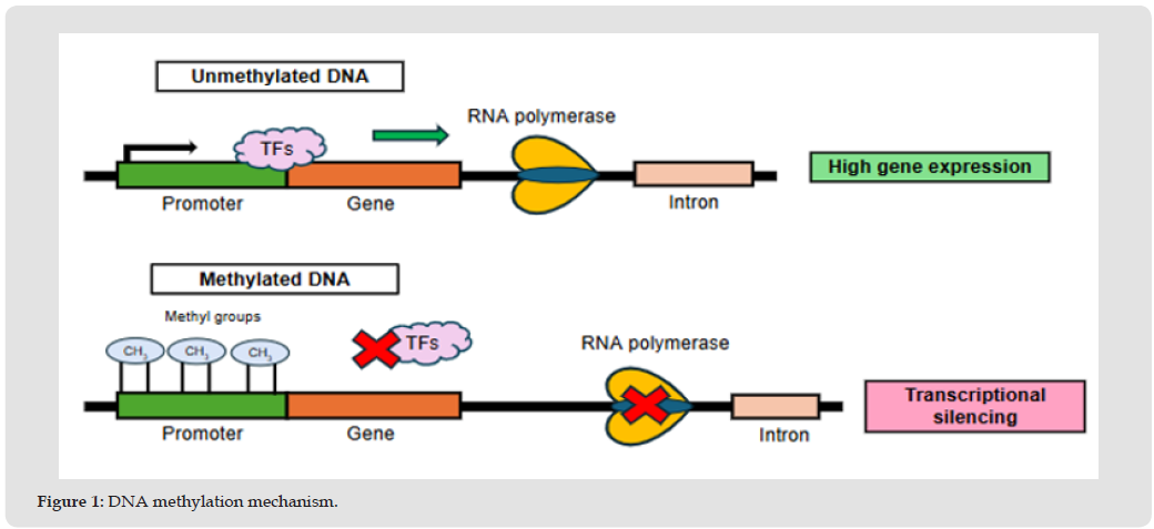
It represents one of the most widely studied epigenetic mechanisms due to its capacity to regulate gene expression without modifying the nucleotide sequence. It consists of appending a methyl group to the carbon 5 of cytosine residues located within CpG dinucleotides via DNA methyltransferases. Generally, methylation of promoter regions is associated with decreased gene transcription, whereas its absence promotes gene expression. Although this mechanism occurs primarily within promoter regions, it can also manifest in exons and introns (Figure 1). In an unmethylated promoter, transcription factors (TFs) and RNA polymerase can bind to the DNA, allowing transcription to start and resulting in high gene expression. In contrast, methylation (CH3) of cytosines in CpG dinucleotides of the promoter prevents transcription factors from being recruited and promotes the establishment of a repressive chromatin state, blocking RNA polymerase action and causing transcriptional silencing. Notably, the relationship between methylation and gene expression depends on the specific DNA region where it occurs. In particular, exonic methylation can be associated with an increase in gene expression, contrasting with what is habitually observed in promoter regions [10].
Figure 1
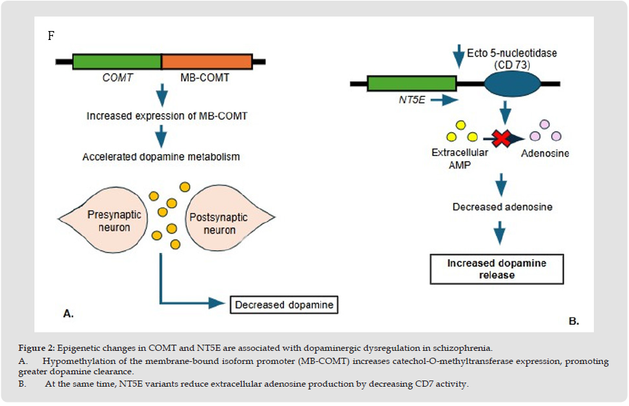
Among the most extensively studied genes is COMT, which encodes catechol-O-methyltransferase, the enzyme responsible for dopamine metabolism in the prefrontal cortex [11]. Khavari & Cairns [12] described hypomethylation in the promoter region of the membrane- bound isoform of this enzyme, which upregulates its expression and, consequently, accelerates dopaminergic metabolism at the frontal lobe level (Figure 2A) [12]. This epigenetic alteration can be linked to the findings described by Peng, et al. [13], who observed a decrease in adenosine availability—a molecule involved in modulating the release of neurotransmitters such as dopamine and glutamate. The reduction in adenosine levels promotes hyperdopaminergic states and disruptions in glutamatergic neurotransmission. The authors attribute this decrease to polymorphisms within the NT5E gene, which encodes the ecto-5’-nucleotidase (CD73) enzyme responsible for converting extracellular AMP into adenosine (Figure 2B) [13]. Another significant gene is GAD1, which produces glutamate decarboxylase (GAD67), thereby facilitating the production of the inhibitory neurotransmitter GABA. Hypermethylation of its promoter region has been identified, a phenomenon associated with GABA depletion and impairments in motor, cognitive, and behavioral functions [14].
Figure 2
Experimental studies demonstrate that maternal immune activation can induce GAD1 hypermethylation and reduce its messenger RNA levels, suggesting that environmental factors during development can alter the regulation of genes involved in neuronal function [12]. Reelin is a glycoprotein secreted by Cajal-Retzius cells during brain development and, subsequently, by GABAergic neurons in the hippocampus and cerebellum of the adult brain. It promotes neuronal migration, cortical stratification, and the maintenance of synaptic plasticity. Its action is exerted by binding to receptors such as the apolipoprotein E receptor 2 (ApoER2) and the very-low-density lipoprotein receptor, whose activation induces the intracellular phosphorylation of the Disabled-1 (Dab1) protein, thereby regulating the organization of the actin and microtubule cytoskeleton [15,16]. Research conducted by Costa, et al. [17] reports a downregulation of this protein due to hypermethylation of the RELN gene at its promoter region [17]. Likewise, Ruzicka, et al. [17] mention that this hypermethylation is predominantly exhibited within GABAergic neurons at the prefrontal cortex level [18]. The functional relevance of this alteration has been demonstrated in animal models, where homozygous reelin-deficient mice display abnormal interneuron positioning in the neocortex, enhanced cognitive impairment, decreased working memory, and stereotyped behavior. Furthermore, they exhibit a decrease in GAD67 mRNA within the prefrontal cortex.
Conversely, mice heterozygous for this gene show reduced dendritic spine density and deficits in prefrontal cortex-dependent cognitive skills, such as reversal learning and fear extinction recall [16]. Decreased reelin expression disrupts neuronal cytoskeleton organization, synaptic connectivity formation, and proteins related to the extracellular matrix and cell adhesion. Among these proteins, secretogranin II stands out; encoded by the SCG2 gene, it is a protein involved in the regulation of secretory granules and neuropeptide packaging. Its downregulation is associated with alterations in synaptic plasticity and cognitive deficits dependent on the dorsolateral prefrontal cortex, particularly in working memory and executive function. Similarly, reduced expression of the L1CAM protein alters cell adhesion and the organization of cortical neuronal networks [19]. Another protein involved is JAM3, a component of endothelial tight junctions. Its disruption can increase vascular barrier permeability and promote processes related to brain damage and neuroinflammation [20]. In the same manner, hypermethylation of serotonergic genes has been identified—particularly HTR1A, HTR2A, and 5-HTT—leading to a decrease in serotonergic neurotransmission (Figure 3) [2].
Figure 3
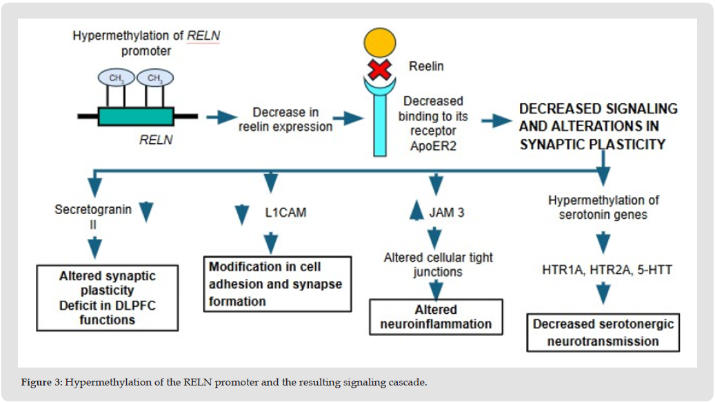
This initial process leads to a reduction in reelin expression and decreases its binding to the ApoER2 receptor, causing defective cell signaling and changes in synaptic plasticity. From this point, four pathways of pathological impact branch out. Delphin, et al. [14] described that numerous genes involved in neuronal growth, cellular differentiation, and the myelination process are hypermethylated in the prefrontal cortex and hippocampus [14]. These alterations could help explain the anomalies observed in neurodevelopmental regulatory genes, such as HUWE1, whose dysfunction affects neural progenitor cell differentiation, a condition associated with intellectual disability and neurodevelopmental disorders [21]. Finally, several studies have demonstrated alterations in the methylation patterns of genes belonging to multiple neurotransmission systems, including MARLIN-1, KCNJ6, HELT, DRD2, and NR3B, which are implicated in regulating GABAergic, glutamatergic, and dopaminergic neurotransmission, as well as NMDA receptor function.
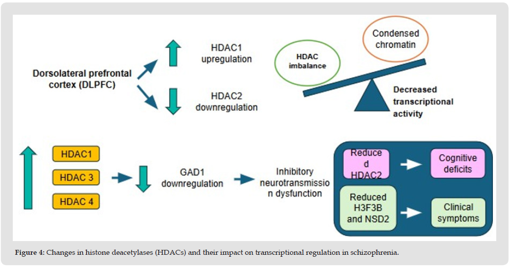
They regulate gene expression through chemical alterations occurring primarily at the amino acid residues of the N-terminal tails of histones H3 and H4, modifying chromatin structure and DNA accessibility to transcription factors without altering the genome nucleotide sequence. They participate in essential processes such as transcriptional regulation, cellular differentiation, DNA repair, and neuronal development [22]. Among the most extensively studied modifications is histone acetylation, a process catalyzed by histone acetyltransferases (HATs) that promotes chromatin relaxation and gene transcription activation. Conversely, histone deacetylases (HDACs) remove these acetyl groups, promoting chromatin condensation and transcriptional repression [23]. Studies conducted on post-mortem brain tissue have demonstrated an increase in cortical levels of histone deacetylase 1 (HDAC1), accompanied by a decrease in histone deacetylase 2 (HDAC2) expression, suggesting an imbalance in the epigenetic mechanisms that control chromatin accessibility and transcriptional activity [17]. These findings have been corroborated by Jiménez Trejo, et al. [24], who described a significant decrease in HDAC2 and an increase in HDAC1 within the dorsolateral prefrontal cortex. The alteration in the relative expression of these enzymes promotes a more condensed chromatin state and downregulates transcriptional activity in genes essential for neuronal function.
Furthermore, they report a relationship between histone deacetylases and the regulation of the GABAergic system, showing a negative correlation between GAD1 expression and the HDAC1, HDAC3, and HDAC4 genes, thereby indicating that increased expression of these enzymes is associated with GAD1 downregulation [24]. Consistently, previous studies conducted by Sharma et al. demonstrated a negative association between GAD1 expression and increased HDAC1 levels, supporting the hypothesis that histone deacetylase-mediated epigenetic alterations contribute to inhibitory neurotransmission dysfunction [12]. Liu W (2021) mention that the histone modification-related genes H3F3B and NSD2 are associated with the clinical symptoms of schizophrenia; additionally, Ganguly S et al. (2021) link histone acetylation to cognitive deficits. Significantly reduced levels of HDAC2 were found in the dorsolateral prefrontal cortex in a post-mortem brain study. Schroeder FA et al. (2021) describe a decrease in HDAC2 within the dorsolateral prefrontal cortex of post-mortem patients (Figure 4) [25]. Changes in the expression of HDAC1, HDAC3, and HDAC4 alter the balance of histone acetylation in the dorsolateral prefrontal cortex, which reduces the expression of genes involved in inhibitory neurotransmission, like GAD1.
Figure 4
They consist of covalent chemical alterations that occur in proteins after their synthesis and constitute a fundamental mechanism for expanding the functional diversity of the proteome. These modifications generate differences in protein structure, stability, subcellular localization, enzymatic activity, and molecular interactions [26]. Within the field of schizophrenia, glycosylation is the most extensively studied post-translational modification; however, ubiquitination and phosphorylation have also been described [27]. N-glycosylation is a process wherein the oligosaccharide N-acetylglucosamine (GlcNAc), mediated by glycosyltransferases, binds to proteins in the endoplasmic reticulum, producing three types of N-glycans: complex, hybrid, and high-mannose polysaccharides. It plays an essential role in protein folding by facilitating the interaction of glycoproteins with the lectin-dependent chaperones calnexin and calreticulin, which ensure they maintain an appropriate conformation for subsequent processing [28]. AMPA receptors are an example of proteins linked to this process, where the GluA2 and GluA4 subunits are the most affected within the dorsolateral prefrontal cortex. Specifically in GluA2, glycans rich in the monosaccharide mannose decrease, indicating abnormal processing of this subunit.
In patients with this pathology, the proportion of unedited GluA2 increases, causing it to prematurely escape the endoplasmic reticulum and give rise to an immature molecule [28,29]. Similarly, excitatory amino acid transporters (EAATs), responsible for glutamate uptake from the synaptic cleft and extracellular space into glial cells, exhibit alterations in their N-glycosylation. There is a decrease in N-glycans in EAAT1 within the anterior cingulate cortex and EAAT2 in the dorsolateral prefrontal cortex, leading to impaired glutamate uptake [28,29]. Likewise, alterations have been identified in the N-glycosylation of the α1, β1, and β2 subunits of the GABA type A (𝐺𝐴𝐵𝐴𝐴) receptor. The density of 𝐺𝐴𝐵𝐴𝐴within the left superior temporal gyrus is increased, whereas the expression of the β1 subunit is decreased at the cerebellar level. Molecularly, this receptor exhibits an increase in mannose residues within its extramembrane domain, suggesting alterations in the processing and maturation of proteins involved in inhibitory neurotransmission [28,29]. Consistent with the alterations observed in this described process, several studies have reported changes in key enzymes involved in N-glycan branching and elongation. Kippe, et al. [30] identified a decrease in the levels of the N-acetylglucosaminyltransferases MGAT4A and B3GNT8 in the dorsolateral prefrontal cortex. The MGAT4A enzyme catalyzes the transfer of N-acetylglucosamine residues to glycans, promoting the formation of complex tri- and tetra-antennary structures.
Meanwhile, B3GNT8 is a polylactosamine synthase enzyme that participates in N-glycan elongation and modification by forming branched lactosamine chains. The decrease in MGAT4A can generate dysregulated binding of lectins, which participate in glycoprotein folding and intracellular trafficking. Furthermore, B3GNT8 interacts with the ubiquitin C protein, which is implicated in processes such as endoplasmic reticulum-associated degradation, DNA repair, and lysosomal degradation. The alteration of these enzymes can compromise protein folding, modify intracellular transport, and affect transmembrane protein internalization, contributing to aberrant glycosylation and cellular dysfunction [30]. Available evidence indicates that alterations in N-glycosylation and its participating enzymes constitute a relevant molecular mechanism in the pathophysiology of schizophrenia by affecting the folding, maturation, intracellular trafficking, and function of proteins essential for neurotransmission. The described changes in glutamatergic receptors, glutamate transporters, and GABAergic receptors suggest that the disruption of this post-translational pathway could contribute to the imbalance between excitatory and inhibitory neurotransmission, prompting alterations in synaptic function and the regulation of neuronal circuits involved in the disease.
In addition to specific alterations in N-glycosylation, changes have been detected in the cellular mechanisms responsible for monitoring proper protein folding and processing in the endoplasmic reticulum. Kim, et al. [31] observed an increase in the expression of the enzyme UDP-glucose/glycoprotein glucosyltransferase 2 (UGGT2) in the dorsolateral prefrontal cortex. This enzyme plays an essential role as a quality control mechanism during protein folding in the endoplasmic reticulum. Additionally, the authors reported an increase in the expression of the synoviolin 1 (SYVN1) complex, an E3 ubiquitin ligase anchored to the endoplasmic reticulum membrane that participates in identifying and tagging misfolded proteins for subsequent degradation via the proteasome system. These findings suggest alterations in protein homeostasis, characterized by an increase in endoplasmic reticulum-associated degradation, which could contribute to the dysfunction of proteins essential for neurotransmission and neuronal operation [31]. In alignment with these alterations in proteostasis and protein quality control mechanisms, Ahmed, et al. [32] highlight the alteration of the DISC1 protein, which is involved in cellular proliferation, differentiation, and migration processes. Truncating mutations or alterations in its gene promote the formation of protein aggregates with cytotoxic effects that disrupt dopaminergic signaling, the cAMP-mediated signaling pathway, and cytoskeletal organization, thereby impacting synaptic function and neurodevelopmental processes.
Another protein of relevance is the neuronal nitric oxide synthase 1 adaptor protein (NOS1AP), a cytosolic protein that interacts with neuronal nitric oxide synthase (nNOS) and participates in regulating nitric oxide-mediated signaling, synaptic plasticity, and neuronal development. An increase in its expression has been reported in the dorsolateral prefrontal cortex and within the dorsal hippocampus of mice, where it produces impairments in social and working memory. Finally, dysbindin-1, encoded by the DTNBP1 gene, plays a fundamental role in maintaining synaptic structure and regulating dopaminergic and glutamatergic systems. Studies in animal models demonstrate that dysbindin-1 overexpression produces impairments in working memory and social interactions—behavioral phenotypes related to schizophrenia. Likewise, it has been observed that specific intronic variants of its gene can modify the alternative splicing process, promoting an increase in the expression of the dysbindin-1B isoform. Collectively, these alterations demonstrate the importance of defects in protein folding, processing, and modification in the pathophysiology of this disease [32].
It is a biological process that occurs following DNA transcription, enabling the generation of distinct RNA variants from a single precursor molecule. It is fundamental for regulating gene expression and expanding the functional diversity of the generated proteome. To achieve this vast diversity, alternative splicing employs various molecular strategies, such as the use of alternative first or last exons, exon skipping, alteration of splice sites, mutually exclusive exon selection, or intron retention. As a result of these mechanisms, a single gene possesses the capacity to encode proteins with entirely different functions. This regulatory role takes on even greater significance within the central nervous system, where alternative splicing is indispensable for neurodevelopment, aging, and the maintenance of essential neuronal functions [33]. Wu, et al. [34] identified aberrant transcript processing patterns in the DCLK1 and PLP1 genes within the superior temporal gyrus. The DCLK1 gene, implicated in neuronal migration, cytoskeletal organization, and synaptic plasticity, exhibited a preferential usage of the TSSIII promoter, driving the expression of isoforms associated with cell survival mechanisms and neuronal remodeling. Similarly, PLP1—one of the primary genes encoding myelin proteins—showed splicing alterations in the PLP and DM20 isoforms, suggesting that post-transcriptional processing defects may contribute to both neurodevelopmental abnormalities and oligodendroglial dysfunction [34].
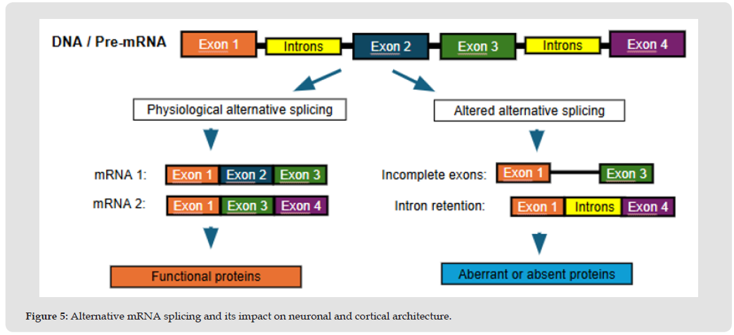
Subsequently, Cohen, et al. [35] analyzed post-mortem samples from the prefrontal cortex and caudate nucleus, identifying multiple differential splicing events in genes associated with neuronal development and synaptic organization. Prominent among the affected genes were ENAH, involved in regulating the actin cytoskeleton and axonal growth, and CPNE3, involved in calcium-dependent intracellular signalling processes. The authors propose that alterations in alternative splicing can modify neuronal architecture and contribute to cortical dysfunction (Figure 5) [35]. From a pre-mRNA, normal post-transcriptional processing produces various protein isoforms essential for synaptic plasticity, myelination, and cytoskeleton dynamics. In contrast, abnormal splicing patterns in key genes like DCLK1, PLP1, and CPNE3 lead to an imbalance in their functional variants. This defect compromises cellular architecture and underlies the structural and cortical dysfunction observed in areas such as the superior temporal gyrus and the prefrontal cortex. Subsequent studies demonstrate that numerous susceptibility genes for this disease generate multiple isoforms with distinct functional properties, capable of modifying essential processes such as neurodevelopment, neurotransmission, and synaptic plasticity [23].
One of the most relevant examples is DISC1, whose regulation via alternative splicing generates diverse isoforms, some of which feature truncations or the loss of critical functional domains. These alterations impair the capacity of DISC1 to interact with intracellular regulatory proteins, thereby compromising processes such as cortical neuronal migration, dendritic arborization, and synaptic plasticity. Furthermore, they modify the activity of fundamental intracellular signaling pathways, including PI3K/AKT/mTOR and GSK3β. Another gene of particular relevance is DTNBP1, responsible for encoding the dysbindin-1 protein. Both Zhang, et al. [23] and Ahmed, et al. [32] have described that alterations in the relative expression of its different isoforms—especially the upregulation of the dysbindin-1B isoform—promote the formation of intracellular aggregates and disrupt synaptic function. These modifications may contribute to characteristic cognitive impairment and working memory deficits [23,32]. The dopamine D2 receptor, encoded by the DRD2 gene, generates two main isoforms through alternative splicing: the short isoform (D2S), localized predominantly at presynaptic terminals and responsible for regulating dopamine release, and the long isoform (D2L), whose function unfolds primarily at the postsynaptic level.
Figure 5
Alterations in the physiological ratio between both isoforms could simultaneously contribute to mesolimbic dopaminergic hyperactivity and cortical dopaminergic hypofunction, providing a molecular mechanism capable of explaining the coexistence of positive symptoms, negative symptoms, and cognitive deficits [23]. North, et al. [36] demonstrate that neuroinflammation modifies transcriptomic processing in brain regions implicated in neuronal generation and maturation. They identified an increase in the expression of COL4A1 and COL4A2, collagen genes participating in the formation of the cerebral basement membrane. This suggests that neuroinflammation could induce fibrosis within the cerebral neurogenic niche, modifying the physical and functional properties of the environment where new neurons are generated. They also detected alterations in genes related to angiogenesis and vascular remodelling, including an overexpression of vascular endothelial growth factor (VEGFA) and its receptors FLT1 and FLT4, accompanied by increased expression of endothelial and vascular smooth muscle cell markers. These results suggest the existence of an active cerebral vascular remodelling process in patients presenting with high inflammation levels.
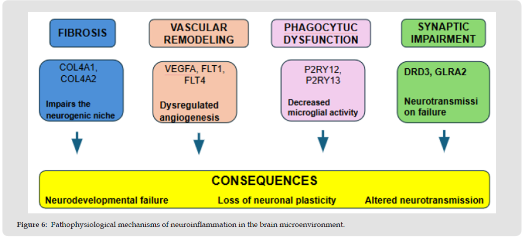
The authors identified a decrease in the expression of genes related to physiological microglial activation and phagocytosis, such as P2RY12 and P2RY13, along with alterations in genes involved in neurotransmission, including DRD3 and GLRA2. These findings suggest that neuroinflammation not only affects neurodevelopment and neuronal plasticity but also the regulation of dopaminergic and glycinergic neurotransmission (Figure 6) [36]. Sustained inflammation alters the regional transcriptomic profile through four converging axes: basal membrane and neurogenic niche fibrosis (COL4A1/2), reactive angiogenesis (VEGFA/FLT1/4), impaired microglial surveillance and phagocytosis (P2RY12/13), and dysregulation of dopaminergic and glycinergic signaling (DRD3, GLRA2). Together, these disturbances compromise the brain’s physical architecture, neurodevelopment, and synaptic plasticity.
Figure 6
This process is part of the final stage of gene expression and is critical for correct neuronal structure and function. It initiates in the ribosomes through mRNA translation, which is divided into the initiation, elongation, termination, and recycling phases. Translational initiation control represents the primary checkpoint of translation and depends on factors such as elF2, elF4E, elF4G, and the elF4F complex, which determine the efficiency with which the ribosome recognizes the mRNA and initiates protein synthesis. In neurons, local translation in dendrites and synaptic spines allows for the rapid production of proteins required for synaptic plasticity, learning, and memory [37]. Mekiten, et al. [38] detected a coordinated overexpression of several genes encoding ribosomal proteins, accompanied by increased expression of HIF1A and NQO1 proteins-findings that suggest a compensatory response to oxidative stress and translational dysfunction. The authors propose that these alterations modify neuronal protein homeostasis and promote an imbalance between excitatory and inhibitory neurotransmission [38]. On the other hand, English, et al. [39] identified a deregulation of the elF2a, elF4, and mTOR pathways, accompanied by modifications in ribosomal protein expression and a global alteration of translation.
Concurrently, in patient-derived olfactory cells, they demonstrated a significant reduction in the rate of protein synthesis, as well as a decrease in seventeen ribosomal proteins. Integrated analysis of proteomics, transcriptomics, and genome-wide association studies identified ElF2AK2 and the elF2/elF4/mTOR regulatory complexes as core components of this molecular alteration [39]. More recent evidence also demonstrates that genes associated with schizophrenia risk are enriched in the local synaptic translatome; that is, the subset of messenger RNAs translated directly in dendrites and synaptic terminals. Gachechiladze and colleagues (2024) identified that numerous susceptibility genes encode proteins whose translation occurs locally at the synapse and which participate in the organization of the postsynaptic density, glutamatergic neurotransmission, and neuronal plasticity. The disruption of local translation constitutes a molecular mechanism capable of contributing to the connectivity and synaptic function alterations observed in this disease [40].
Intracellular signaling pathways play a fundamental role in regulating cell growth, proliferation, differentiation, and survival. The PI3K/AKT/mTOR pathway has acquired special relevance in the study of schizophrenia due to its involvement in processes essential for brain functioning, including neurodevelopment, synaptic plasticity, protein synthesis, and long-term memory consolidation. Protein kinase B (AKT) and mammalian target of rapamycin (mTOR) are serine/ threonine kinases that act as central nodes of multiple cellular signaling networks. AKT activation is regulated by growth factors, cytokines, and neurotransmitters, whereas mTOR integrates metabolic and trophic signals to coordinate the cellular response to various physiological stimuli. Once activated, mTOR forms two functionally distinct multiprotein complexes: the mTORC1 complex, primarily responsible for regulating ribosomal biogenesis, protein synthesis, and autophagy; and the mTORC2 complex, involved in actin cytoskeleton organization, cell survival, and the regulation of AKT activity. Due to these functions, disruption of the PI3K/AKT/mTOR pathway can simultaneously affect neuronal architecture, brain connectivity, and synaptic plasticity. The study conducted by Chadha and colleagues (2021) shows that AKT kinase expression is reduced at the level of the dorsolateral prefrontal cortex, as well as the GβL subunit in mTORC1 and mTORC2.
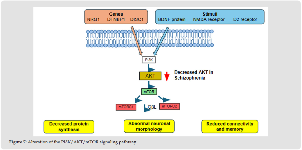
Furthermore, the expression of AKT in its active form (phospho- AKT S473 and phospho-AKT T308) and the phosphorylation state of mTOR were reduced, thereby decreasing the activity of both kinases and resulting in a downregulation of the AKT-mTOR pathway. Studies in mice have demonstrated that AKT deficiency causes alterations in learning and synaptic plasticity at the hippocampal and prefrontal cortex levels. Likewise, mTOR inhibition reduces protein synthesis in hippocampal and cerebral cortex neurons, linking the latter to cognitive deficits. mTOR integrates signaling from NMDA receptors, brain-derived neurotrophic factor (BDNF), and dopaminergic receptors through phosphoinositide 3-kinase (PI3K). The disruption of this pathway generates reduced protein synthesis, abnormal neuronal morphology, and decreased brain network connectivity [9]. The AKT protein occupies a particularly important position within this signaling network, acting as a molecular integrator of multiple neurotransmitter systems. Its activity is co-regulated by D2 dopaminergic receptors, serotonergic receptors, BDNF, and various growth factors, through intermediary molecules such as glycogen synthase kinase-3 beta (GSK-3β) and the β-arrestin-2 protein. Balu and colleagues (2012) demonstrated that mice deficient in the AKT1 isoform present a significant decrease in hippocampal cell proliferation, accompanied by alterations in long-term potentiation in the CA1 region.
Likewise, these animals exhibited deficits in spatial memory, while studies in patients with schizophrenia evidenced alterations in AKT1 signaling in the prefrontal cortex [41]. In addition to its direct participation in neurotransmission and neuronal plasticity, AKT constitutes a convergence point for various susceptibility genes. Prominent among them are NRG1 (neuregulin), DTNBP1 (dysbindin), and DISC1, whose gene products regulate neuronal growth, cell differentiation, synaptic circuit formation, and brain plasticity. The functional convergence of these genes on AKT-mediated signaling suggests that the alteration of this pathway could represent a common pathophysiological mechanism capable of explaining the clinical and molecular heterogeneity characteristic of schizophrenia Figure 7 [41]. AKT kinase acts as a central integrator of multiple extracellular signals (NMDA receptors, dopaminergic receptors, and susceptibility genes like DISC1). In schizophrenia, there is a hypofunction and pathological hypophosphorylation of the AKT-mTOR axis and its complexes (mTORC1/2), which disrupts the regulation of ribosomal biogenesis and the cytoskeleton. This defective cascade physiologically results in poor protein synthesis, changes in neuronal morphology, and deficits in synaptic plasticity and memory.
Figure 7
Reelin protein, whose expression is decreased due to hypermethylation of the RELN gene, induces the recruitment of PI3K and AKT upon binding to the ApoER2 receptor and subsequent phosphorylation of the Dab1 protein. Subsequently, activation of the mTOR cascade promotes phosphorylation of S6K and mediates the trophic effects of reelin on hippocampal growth and dendritic arborization— processes that are fundamental for the maintenance of synaptic plasticity and neuronal connectivity [15]. Complementarily, genetic studies have identified primary alterations in components of this signaling pathway; Enriquez Barreto, et al. [42] described a mutation in the promoter region of the PIK3C3 gene, corresponding to class III phosphoinositide 3-kinase, which produces a decrease in its gene transcription in patients with schizophrenia. This finding provides additional evidence that dysregulation of the PI3K/AKT/mTOR pathway can originate from both epigenetic and post-transcriptional mechanisms as well as from direct genetic alterations [42].
Synaptic pruning constitutes an essential physiological process during neuro development, whereby redundant or poorly functional neuronal connections are eliminated to optimize the organization and efficiency of brain circuits. This mechanism depends on a highly regulated interaction between neurons, microglia, and components of the immune system. Evidence accumulated over recent decades suggests that alterations in synaptic pruning and neuroinflammatory processes play a central role in the pathophysiology of schizophrenia, contributing to the disruptions in brain connectivity, neuronal plasticity, and cognitive functioning observed in this disorder. One of the genes involved is PLA2G4A, which encodes cytosolic phospholipase A2 (cPLA2), a participant in membrane phospholipid metabolism and the production of inflammatory mediators derived from arachidonic acid. Gao, et al. [43] demonstrated that deletion of this gene decreases cPLA2 levels and increases susceptibility to schizophrenia, whereas Yang, et al. [25] found increased PLA2G4A transcription and higher cPLA2 activity in patients with the disease [25, 44]. These alterations may be linked to a dysregulation in synaptic plasticity and neuronal metabolism, as membrane phospholipids are essential for the maintenance of synaptic receptors, neurotransmitter release, and the structural stability of dendritic spines.
Therefore, PLA2G4A alterations could promote synaptic vulnerability and facilitate the action of pathological mechanisms implicated in schizophrenia, such as synaptic pruning and neuroinflammation [25, 43]. The identification of the complement system’s involvement in regulating synaptic pruning has been achieved thanks to research conducted by Sekar et al. (2025), demonstrating that structural variations in the C4 gene, especially those associated with increased expression of the C4A isoform, significantly increase the risk of developing schizophrenia due to excessive activation of synaptic pruning during neurodevelopment [2]. Subsequently, Gu et al. confirmed that alleles associated with higher brain expression of this gene correlate with elevated C4 protein levels and a reduction in cortical thickness [44]. The information presented by Phadke, et al. [45] expands on this pathophysiological mechanism by demonstrating that increased C4A activity produces dendritic spine loss independently of the classical pathway mediated by the microglial CR3 receptor. Furthermore, they identified that C4 activation alters the intracellular trafficking of AMPA receptors, particularly those containing the GluR1 subunit, decreasing their recycling to the postsynaptic membrane. This observation can be directly linked to alterations in synaptic plasticity, as the reduction of functional AMPA receptors compromises long-term potentiation, memory, and learning, contributing to characteristic cognitive deficits [45]. Likewise, the increase in C4A expression observed by Mou et al. (2021), particularly in the brain’s subventricular zone, establishes an important relationship between the complement system and neurodevelopment [2]. This region contains neural progenitor cells and actively participates in neurogenesis processes. Therefore, C4A overexpression could contribute to the described neurodevelopmental alterations, those related to genes such as HUWE1, whose function is essential for the proliferation and differentiation of neural progenitor cells [21]. Another important aspect is the negative regulation of the complement system mediated by the CSMD1 gene. The findings of Hatzimanolis et al. demonstrated that patients with a first psychotic episode present decreased CSMD1 expression and increased serum levels of C4A, favoring a state of complement hyperactivation. The reduction in CSMD1 expression was additionally associated with working memory alterations and impairment of executive functions dependent on the prefrontal cortex. These findings suggest that the loss of physiological complement regulation amplifies the pathological activity of C4A, contributing to neuroinflammation and the excessive elimination of neuronal connections [46].
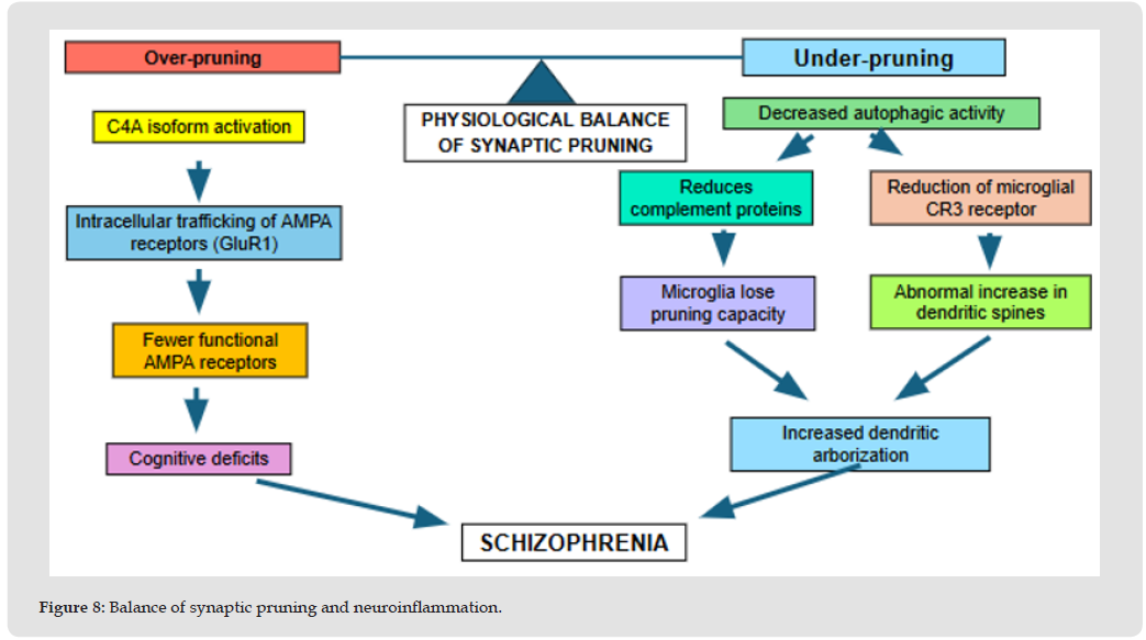
The involvement of the immune system in schizophrenia is also supported by the findings of Savukoski, et al. [47], who identified elevated concentrations of the soluble terminal complement complex in peripheral blood. These results suggest the presence of persistent immune activation, which could favor microglial activation and perpetuate neuroinflammatory processes, including the participation of genes such as CTSS, involved in microglial activation and pathological synaptic remodeling [47]. On the other hand, the findings of Su, et al. [48] link the complement system to alterations in autophagy. They demonstrated that decreased autophagic activity during critical periods of brain development significantly reduces the expression of complement proteins, including C1q, C3b, and C4, as well as the expression of the microglial CR3 receptor. Consequently, microglia lose their capacity to recognize and eliminate surplus synapses, producing an abnormal increase in dendritic spines and an aberrant organization of neuronal circuits [48]. These findings are particularly relevant because they suggest that both excessive and defective synaptic pruning can contribute to the pathophysiology of schizophrenia.
While C4A overexpression promotes excessive elimination of neuronal connections, decreased autophagy generates insufficient synapse elimination, equally disrupting the functional organization of brain circuits Figure 8 [2,48]. The disease arises when the homeostatic balance is broken, either due to excessive pruning (caused by overexpression of C4A and loss of functional AMPA receptors) or defective pruning (linked to microglial dysfunction and reduced autophagy, leading to abnormal retention of dendritic spines).
Figure 8
The reviewed evidence allows for the understanding that the pathophysiology of schizophrenia is the result of a complex interaction among genetic, epigenetic, protein, and cellular alterations that affect the development and functioning of the central nervous system. DNA methylation mechanisms, histone modifications, post-translational alterations, alternative RNA splicing, and the dysregulation of protein synthesis can modify the expression, processing, stability, and function of proteins essential for neurotransmission and neuronal plasticity. In turn, the alteration of the PI3K/AKT/mTOR signaling pathway, as well as processes such as synaptic pruning and neuroinflammation, contribute to the dysfunction of neuronal circuits involved in neurodevelopment, cognition, and the regulation of dopaminergic, glutamatergic, and GABAergic neurotransmission. Taken together, these mechanisms suggest that the disease cannot be explained by an isolated alteration of a single gene, neurotransmitter, or brain circuit, but rather as a consequence of the convergence of multiple interacting molecular processes. Their dysregulation can compromise neuronal connectivity, synaptic maturation, the excitatory- inhibitory balance, and the functional adaptive capacity of the brain.
Therefore, the study of these alterations allows for a deeper understanding of the biological and clinical heterogeneity of schizophrenia, as well as the identification of potential mechanisms involved in its development and new opportunities for researching therapeutic strategies targeted at its molecular bases.
We declare that we have no conflict of interest.

International Association of Landscape Archaeology, Czech Glass Society, Czech Republic

Department of Chemistry, Semenov Institute of Chemical Physics, USSR Academy of Sciences, Moscow, Russia

Associate Professor at Department of Breast and Thyorid Surgey, Chongqing General Hospital, China

Professor of Nuclear Medicine, Faculty of Medicine and Surgery, University of Milan, Milan, Italy

Department of Pharmaceutical Sciences, South University School of Pharmacy, USA





 Abstract
 PDF