[email protected]
+1 (502) 904-2126
One Westbrook Corporate Center, Suite 300, Westchester, IL 60154, USA
Site Map
 ISSN: 2574 -1241
Received: September 22, 2026; Published: October 05, 2026
*Corresponding author: Li Ye, Naxi District People’s Hospital, Luzhou City, Sichuan Province, China
DOI: 10.26717/BJSTR.2026.67.010427
Objective: To investigate the clinical efficacy and complication profile of a novel minimally invasive elastic micro-
motion implant in the treatment of acute inferior tibiofibular joint injuries, thereby providing a basis for
selecting appropriate surgical techniques in clinical practice.
Methods: A total of 72 patients with acute inferior tibiofibular joint injuries admitted between February 2021
and October 2023 were retrospectively enrolled and divided into an observation group (36 patients receiving
fixation with the novel minimally invasive elastic micro-motion implant) and a control group (36 patients receiving
conventional cortical screw rigid fixation). Perioperative parameters, postoperative AOFAS ankle-hindfoot
score, VAS pain score, and complication rates were compared between the two groups. All patients were followed
up for 12–24 months, with a median follow-up duration of 16.5 months.
Results: The operative time in the observation group was longer than that in the control group (P<0.05), while
intraoperative blood loss and incision length were significantly shorter in the observation group compared to
the control group (P<0.05). At the final follow-up, the AOFAS score in the observation group (87.36±5.82) was
higher than that in the control group (81.47±6.35), whereas the VAS score (1.51±0.74) was lower than that in the
control group (2.28±0.91); all differences were statistically significant (P<0.05). The overall complication rate
in the observation group was 8.33%, significantly lower than that in the control group (25.00%) (P<0.05). In
the observation group, complications were predominantly mild soft tissue irritation, whereas the control group
exhibited serious complications such as screw breakage, loss of reduction, and traumatic arthritis. Conclusion:
The novel minimally invasive elastic micro-motion implant for the treatment of acute inferior tibiofibular syndromes
offers minimal trauma, accelerated rehabilitation, and fewer complications; it preserves the physiological
micro-motion of the tibiofibular syndrome, promotes superior long-term ankle joint functional recovery, and
demonstrates higher clinical safety. Therefore, this implant is worthy of widespread clinical application.
Keywords: Acute Inferior Tibiofibular Syndromes; Minimally Invasive Surgery; Elastic Micromotion Implant; Internal Fixation; Complications; Ankle Joint Injury
Acute inferior tibiofibular syndesmosis injury is a common type of ankle joint injury in clinical practice, often occurring in conjunction with ankle fractures or high-grade sprains. If reduction and fixation are inadequate, it can readily lead to chronic ankle instability, persistent pain, and post-traumatic arthritis, significantly impairing patients’ quality of life and ability to engage in physical activity. Conventional rigid fixation with cortical screws can achieve anatomical reduction; however, it completely restricts the physiological micromotion of the tibiofibular syndesmosis, which contradicts the biomechanical characteristics of the ankle joint. Under prolonged mechanical stress, this approach may result in screw breakage and loss of reduction, necessitating secondary surgery for implant removal— thereby increasing both the healthcare burden and surgical trauma for patients [1]. Modern biomechanical studies have demonstrated that during ankle joint motion, the tibiofibular syndesmosis exhibits physiological micromotion ranging from 1–2 mm [2]. An ideal internal fixation system should therefore balance reduction stability with physiological range of motion.
The novel minimally invasive elastic micromotion implant employs a cable-and-gasket dynamic fixation mechanism, enabling the maintenance of anatomical reduction while preserving physiological micromotion of the syndesmosis. Furthermore, this technique is performed via a minimally invasive approach, resulting in reduced tissue trauma and aligning with the principles of enhanced recovery after surgery (ERAS) [3]. Although this procedure has been clinically applied for a relatively short period, its long-term efficacy and complication profiles still require further systematic evaluation. This study conducts a comparative analysis of the clinical outcomes of these two fixation modalities to systematically assess the clinical utility and safety risks associated with the novel elastic implant, thereby providing a reference for standardized clinical practice.
Subject Investigated
A total of 72 patients with acute distal tibiofibular syndesmotic injury admitted to our hospital between February 2021 and October 2023 were enrolled; all cases were confirmed by X-ray or CT imaging, and the time interval from injury to surgery was less than 3 weeks. Inclusion criteria: confirmed diagnosis of acute distal tibiofibular syndesmotic dislocation with instability; complete clinical and follow-up data; follow-up duration ≥12 months. Exclusion criteria: chronic injuries, prior history of ankle surgery, severe osteoarthritis, open fractures, or patients unable to comply with follow-up visits. Patients were divided into two groups based on the surgical approach, with 36 cases in each group. The observation group comprised 21 males and 15 females, with a mean age of (38.42 ± 8.71) years; the control group comprised 23 males and 13 females, with a mean age of (39.17 ± 9.03) years. There were no statistically significant differences between the two groups in terms of gender, age, mechanism of injury, or baseline preoperative clinical characteristics (P> 0.05), indicating comparability between the groups. This study was approved by the Institutional Review Board of our hospital, and all patients provided written informed consent.
Surgical Procedure
Observation Group: Novel minimally invasive elastic micro-motion implant fixation. The patient assumes a supine position, undergoes intraspinal anesthesia, and is placed under tourniquet control. Following routine management of concomitant fractures, a minimally invasive incision (1.5–2.0 cm) is made on the lateral aspect of the distal fibula, with careful protection of the superficial peroneal nerve. Under fluoroscopic guidance, a fibular-to-tibial bone tunnel is created 2–3 cm above the ankle joint surface; an elastic micromotion cable and bilateral anchoring pads are then implanted. The cable tension is finely adjusted, and a stress test is performed under fluoroscopy to confirm satisfactory anatomical reduction of the distal tibiofibular syndesmosis [4]. The incision is irrigated and sutured, with no need for drainage or cast immobilization.
Control Group: Conventional cortical screw rigid fixation. The anesthesia and positioning were identical to those in the observation group; a 3–5 cm lateral incision was made to fully expose the distal tibiofibular joint. After direct visualization of the reduction, a four-layer cortical rigid fixation was performed using 3.5 mm cortical screws. Following fluoroscopic confirmation of satisfactory reduction, a drainage tube was placed, and the wound was sutured in layers. Postoperative short-leg plaster cast immobilization was applied for 4–6 weeks; the screws were removed during a secondary surgical procedure 3–4 months after the initial surgery [5].
Postoperative Management
Both groups received routine postoperative analgesic, edema-reducing, and prophylactic anti-infective therapy. The observation group commenced ankle pump exercise training on the day following surgery; partial weight-bearing was gradually introduced after 2 weeks, and full weight-bearing was achieved between 4–6 weeks postoperatively. The control group underwent only toe mobilization during the cast immobilization period; after cast removal, functional exercises were progressively initiated, and full weight-bearing was avoided for the first 3 months postoperatively. All patients underwent regular follow-up radiographic examinations at 1, 3, 6, and 12 months postoperatively to assess reduction status, implant position, and joint degeneration.
Observation Indicators
Collect perioperative clinical parameters (including operative time, intraoperative blood loss, and incision length) for both groups; assess preoperative and final follow-up ankle joint function and pain severity using the AOFAS Ankle–Heel Score and VAS pain score; perform imaging evaluation to determine the occurrence of reduction loss; document the incidence of complications such as incisional infection, internal fixation failure, soft tissue irritation, and traumatic arthritis.
Statistical Analysis
Data were analyzed using SPSS 26.0 statistical software. Measurement data were expressed as mean ± standard deviation; intergroup comparisons were performed using the independent samples t-test. Categorical data were expressed as percentages (%); comparisons were conducted using the χ² test. A P-value <0.05 was considered statistically significant.
Comparison of Perioperative Clinical Parameters Between the Two Groups
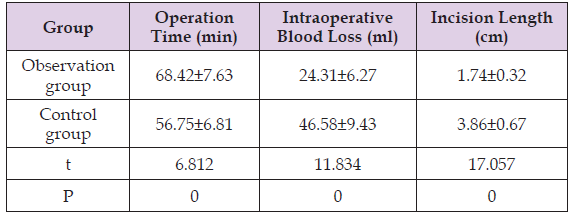
In the observation group, due to the need for precise localization of the bone tunnel and fine adjustment of cable tension, the surgical procedures were more meticulous, resulting in a significantly longer operative time compared to the control group (P<0.05). However, the minimally invasive approach in the observation group eliminated extensive soft tissue dissection, leading to reduced intraoperative trauma, less blood loss, and a markedly shorter incision length relative to the control group. The perioperative trauma parameters in the observation group were also significantly superior to those in the control group (P<0.001). All procedures were completed successfully in every patient, with no occurrences of vascular or nerve injury or conversion to open surgery; detailed results are presented in Table 1.
Table 1: Comparison of perioperative clinical parameters between the two patient groups.
Ankle Joint Function and Pain Scores
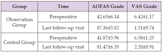
There was no significant difference in AOFAS scores or VAS scores between the two groups before surgery (P> 0.05), indicating baseline equivalence. After surgery, both groups demonstrated significant improvement in symptoms and function; however, the observation group exhibited faster rehabilitation progress and was able to initiate standardized functional training at an earlier stage without significant joint stiffness. At the final follow-up, the observation group showed a significantly higher AOFAS score and a significantly lower VAS pain score, with the intergroup difference being statistically significant (P <0.05), suggesting that elastic fixation is more conducive to ankle joint function recovery and reduction of long-term residual pain; see Table 2 for details.
Table 2: Comparison of AOFAS and VAS scores between the two patient groups before surgery and at the last follow-up visit.
Incidence of Complications
In the observation group, only three cases of mild complications occurred, with a total incidence rate of 8.33%, including two cases of local soft tissue irritation and one case of short-term postoperative swelling; all of these complications completely resolved following conservative intervention. No cases of wound infection, internal fixation failure, loss of reduction, or arthritis occurred, and no second surgeries were required. In the control group, there were nine cases of complications, with a total incidence rate of 25.00%, including wound infection, screw breakage, loss of reduction, and mild traumatic arthritis; most of these were medium-to long-term adverse events. The difference in complication rates between the two groups was statistically significant (χ² = 4.599, P = 0.032), indicating that the novel elastic implant demonstrated superior safety profiles.
The key to treating acute inferior tibiofibular syndesmosis injury lies in achieving precise reduction, stable fixation, and preservation of the normal biomechanical function of the ankle joint. Conventional rigid screw fixation completely restricts tibiofibular micro-motion; during ankle movement, stress concentrates on the internal fixation devices, which can lead to screw fatigue failure, loss of reduction, and necessitate secondary removal procedures—thereby imposing additional trauma and increased medical costs. Furthermore, this approach is associated with a higher incidence of post-traumatic osteoarthritis in the long term. The complication rate of 25.00% observed in the control group of this study further underscores the limitations of traditional surgical techniques. In contrast, the novel minimally invasive elastic micro-motion implant employs a dynamic fixation mode, allowing it to maintain anatomical reduction while preserving physiological tibiofibular micro-motion, thereby fully aligning with the biomechanical characteristics of the ankle joint. Simultaneously, the minimally invasive incision reduces soft tissue and periosteal damage, facilitating early functional rehabilitation and effectively mitigating complications such as joint stiffness and muscle atrophy associated with prolonged cast immobilization.
The results of this study demonstrate that the observation group exhibited less perioperative trauma, superior postoperative functional recovery, milder pain symptoms, and eliminated the need for routine secondary surgeries, demonstrating significant clinical advantages. The only controllable adverse event associated with this technique was mild soft tissue irritation, which was frequently related to superficial placement of the spacer or improper adjustment of cable tension; however, these risks can be effectively minimized through standardized procedures—including precise bone tunnel localization, appropriate spacer positioning, and moderate tension regulation. This study has limitations, including being a single-center investigation with a small sample size and short-term follow-up; the long-term risk of joint degeneration requires further validation through large-sample, long-term follow-up studies [6]. In conclusion, the use of novel minimally invasive elastic micro-motion implants for the treatment of acute distal tibiofibular union injuries offers the advantages of being minimally invasive, safe, and associated with excellent functional recovery; its incidence of complications is significantly lower than that of traditional rigid fixation, aligns with the principles of Enhanced Recovery After Surgery (ERAS), and makes it the preferred surgical approach for the clinical management of acute distal tibiofibular union injuries.
Supported by Luzhou Science and Technology Program (No. 2024SYF189).

International Association of Landscape Archaeology, Czech Glass Society, Czech Republic

Department of Chemistry, Semenov Institute of Chemical Physics, USSR Academy of Sciences, Moscow, Russia

Associate Professor at Department of Breast and Thyorid Surgey, Chongqing General Hospital, China

Professor of Nuclear Medicine, Faculty of Medicine and Surgery, University of Milan, Milan, Italy

Department of Pharmaceutical Sciences, South University School of Pharmacy, USA





 Abstract
 PDF