[email protected]
+1 (502) 904-2126
One Westbrook Corporate Center, Suite 300, Westchester, IL 60154, USA
Site Map
 ISSN: 2574 -1241
ISSN: 2574 -1241
ISSN: 2574 -1241
Received: August 10, 2026; Published: August 25, 2026
*Corresponding author: Yong gang Hu, Department of Clinical Laboratory, People’s Hospital of Naxi District, 219 Renmin East Road, Naxi District, Luzhou City 646300, Sichuan Province, China
DOI: 10.26717/BJSTR.2026.66.010357
Chronic atrophic gastritis and gastric intestinal metaplasia are evaluated through endoscopy, histology and serology, yet intervention studies rarely define how change in the visible mucosal field should be measured. This limitation is evident in studies of folate, vitamin B12 and related regimens, in which histologic scores, symptoms and composite response rates predominate, while endoscopic findings are often reported descriptively. The resulting evidence cannot determine whether an apparent visual improvement represents reduced inflammation, a smaller endoscopic atrophic field, contraction of intestinal metaplasia, histologic stage migration or only measurement variability. In this narrative review, vitamin B intervention studies are used as a methodological test case rather than as proof of efficacy. We distinguish five response domains: inflammatory improvement, reduction in endoscopic atrophy extent, reduction in the endoscopic field of intestinal metaplasia, histologic regression or stage migration, and biomarker concordance. High-definition white-light endoscopy can document global mucosal and atrophic-border features, whereas narrow-band imaging, blue-light imaging and linked color imaging can improve recognition and mapping of intestinal metaplasia. However, endoscopic grading of gastric intestinal metaplasia (EGGIM) and the Kimura-Takemoto classification were developed primarily for cross-sectional staging and risk stratification. Their responsiveness, minimal clinically important change and longitudinal reproducibility remain insufficiently validated. Current vitamin B evidence provides a limited and heterogeneous therapeutic signal, particularly for inflammatory and atrophic outcomes in selected populations, but does not establish reliable reversal of established intestinal metaplasia. Prospective validation is required before endoscopic mucosal response can function as an independent efficacy endpoint.
Keywords: Chronic Atrophic Gastritis; Gastric Intestinal Metaplasia; Endoscopic Endpoint; Folic Acid; Vitamin B12; Image-Enhanced Endoscopy
Abbreviations: CAG: Chronic Atrophic Gastritis; GIM: Gastric Intestinal Metaplasia; HD-WLE: High-Definition White-Light Endoscopy; (BLI): Blue-Light Imaging; LCI: Linked Color Imaging; EGGIM: Endoscopic Grading of Gastric Intestinal Metaplasia
A Brief Synopsis
This review proposes a trial-ready framework for endoscopic mucosal response as a multidomain endpoint in chronic atrophic gastritis, drawing on vitamin B studies.
Chronic atrophic gastritis (CAG) and gastric intestinal metaplasia (GIM) occupy a clinically important position in the gastric precancerous cascade [1,2]. Their relevance is not determined by symptoms, which correlate poorly with cancer risk, but by lesion distribution, severity, etiology and persistence. Contemporary guidance therefore combines high-quality endoscopy with topography-aware histology for diagnosis, staging and surveillance [3-8]. This multimodal approach is established for risk assessment, but not for measuring response to non-eradication interventions. The measurement gap is particularly visible in studies of folate, vitamin B12 and related vitamin B regimens. These studies have reported changes in pathology scores, symptom scales or composite ‘effective rates’, but they rarely specify whether the gastric mucosa became less inflamed, whether the atrophic border changed, or whether the endoscopic field of GIM contracted. As a result, the clinically intuitive question of whether the stomach ‘looks better’ remains methodologically ambiguous. This review argues that visual change should neither be dismissed as subjective nor promoted as a surrogate for histologic reversal. Instead, endoscopic mucosal response should be developed as a domain-specific, longitudinal and reproducible endpoint whose interpretation depends on concordance with mapped histology and, secondarily, serologic biomarkers. Vitamin B intervention studies provide a useful test case because they contain a plausible biological rationale and a modest therapeutic signal, yet also expose the consequences of poorly standardized outcome definitions.
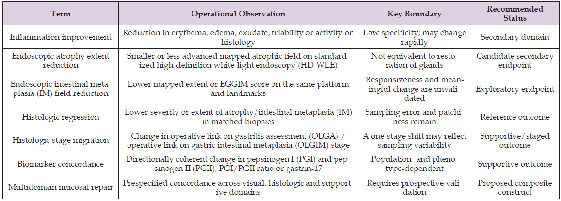
This narrative review prioritizes clinical guidelines, validation studies of endoscopic staging systems, diagnostic-accuracy studies of image-enhanced endoscopy, histologic risk-staging literature, biomarker studies and intervention reports involving folate or vitamin B12. Targeted PubMed searches were updated through 23 June 2026. The review is not a systematic review and does not estimate a pooled treatment effect. Its purpose is methodological: to identify what can currently be measured, what remains unvalidated and how future intervention trials should define change. Terminology is central to this task. ‘Inflammation improvement’, ‘atrophy regression’, ‘intestinal metaplasia regression’, ‘endoscopic field reduction’, ‘histologic stage migration’ and ‘biomarker concordance’ describe different observations. They should not be used as synonyms. We use endoscopic mucosal response as the neutral visual term. Mucosal repair is reserved for a multidomain interpretation in which visual change is supported by histology, biomarkers or both Table 1.
Table 1: Terminology and endpoint hierarchy.
Biological Plausibility Does Not Establish Mucosal Reversal
Folate and cobalamin participate in one-carbon metabolism, nucleotide synthesis and methyl-group transfer [9,10]. Deficiency may coexist with CAG, particularly in corpus-predominant autoimmune disease, where impaired acid and intrinsic-factor physiology can contribute to iron and vitamin B12 deficiency [11]. Genetic associations involving methylenetetrahydrofolate reductase (MTHFR) and epidemiologic associations involving low vitamin B12 provide etiologic context, but they are not treatment-efficacy evidence [12,13]. These mechanisms make correction of deficiency clinically necessary and provide a plausible context for epithelial restitution. They do not, however, demonstrate that supplementation restores lost gastric glands or eliminates established metaplastic clones. The direction of causality also differs across phenotypes. In autoimmune gastritis, vitamin B12 deficiency is often a consequence of oxyntic gland loss; replacement corrects deficiency but is not evidence that the autoimmune atrophic process has reversed. In Helicobacter pylori-associated multifocal atrophy, any observed change may be confounded by eradication, acid-suppressive therapy, diet, baseline folate status or concomitant traditional medicines. Mechanistic plausibility must therefore be separated from treatment efficacy.
The Clinical Signal Is Limited, Heterogeneous and Lesion- Specific
A 2023 study of 96 H. pylori-negative patients compared regimens containing weifuchun alone, weifuchun plus folic acid, or weifuchun plus folic acid and vitamin B12. Histopathologic atrophy improved more often with added folic acid than with weifuchun alone, particularly in less advanced stages, but the small sample, co-interventions, genotype analyses and limited follow-up restrict causal inference about folic acid itself [14]. The study supports a treatment signal in a selected population, not a general claim of reversal. A 2022 meta-analysis reported benefits for gastric precancerous conditions, including atrophy and GIM [15]. Its evidentiary base included regional controlled studies, a conference report and unpublished data, with substantial variation in dose, background therapy, outcome definitions and study quality. The doses described in several included trials also exceed routine nutritional replacement. A 2025 meta-analysis in H. pylori-associated CAG similarly reported favorable composite outcomes, but the underlying studies remained dominated by local practice patterns and heterogeneous regimens [16]. These findings justify further trials while requiring cautious wording such as ‘may improve selected histologic outcomes’ rather than ‘reverses gastric precancerous lesions.
Retrospective reports of multicomponent products combined with other drugs cannot be attributed to vitamin B12 alone [17]. Across this literature, evidence is more credible for improvement in inflammatory activity or atrophy scores than for durable disappearance of established GIM. This lesion hierarchy should determine endpoint timing and language.
Why Conventional Response Measures are Insufficient
Histology is indispensable but spatially sparse. Symptoms are clinically relevant but weakly linked to premalignant burden. Composite effective rates can combine symptoms, endoscopic impressions and pathology into a single proportion, obscuring which domain changed and how it was measured. Narrative endoscopy reports are especially vulnerable to observer expectation, selective photography and inconsistent insufflation or image-enhancement settings. The correct response is not to privilege one modality. Endoscopy surveys a field, biopsy samples points, and serology reflects organ-level physiology. Each modality answers a different question. A trial can exploit this complementarity only when each outcome is defined independently before concordance is assessed.
Domain 1: Inflammatory Normalization
Reduced erythema, edema, adherent mucus, friability or surface irregularity may be visible within months. These features are responsive but nonspecific and sensitive to preparation, insufflation, bile reflux, recent medication and imaging settings. They should be recorded as an inflammatory domain rather than interpreted as atrophy regression.
Domain 2: Change in Endoscopic Atrophy Extent
Atrophy may be suggested by pallor, increased vascular visibility, fold loss and an identifiable atrophic border. A longitudinal endpoint should record the mapped position and extent of that border at fixed landmarks. The preferred term is reduction in endoscopic atrophy extent. ‘Atrophic repair’ should be avoided unless matched histology demonstrates coherent glandular improvement.
Domain 3: Change In the Endoscopic Field of Intestinal Metaplasia
Image-enhanced endoscopy can delineate metaplastic mucosa more accurately than white light alone [18-26]. A smaller visible field may represent true contraction, altered detectability, incomplete remapping or observer variability. Endoscopic IM field reduction is therefore a descriptive endpoint. Histologic IM regression requires matched biopsy evidence, while complete reversal is a substantially stronger claim that current vitamin B evidence does not establish.
High-Definition White-Light Endoscopy
High-definition white-light endoscopy (HD-WLE) remains the baseline examination because it documents preparation quality, anatomy, folds, vascular visibility, the atrophic border and focal lesions. Longitudinal use requires a predefined photographic map, comparable insufflation, image orientation and cleaning. Without these controls, apparent border movement can arise from technique rather than biology.
NBI, BLI and LCI
Narrow-band imaging (NBI) has the largest evidence base for identifying GIM, including metaplasia-associated surface and color patterns such as the light blue crest [19-22]. Earlier dye-based magnification chromoendoscopy established that metaplastic fields can be classified reproducibly under enhanced visualization [27]. Bluelight imaging (BLI) and linked color imaging (LCI) can also improve contrast and field delineation [28-32]. These technologies support detection and mapping, but diagnostic accuracy is not the same as responsiveness to change. Device generation, magnification, processor settings, operator training and the selected color-sign definition can all affect longitudinal comparability.
EGGIM: Suitable for Staging, not Yet Validated as a Treatment- Response Scale
Endoscopic grading of gastric intestinal metaplasia (EGGIM) scores the endoscopic extent of GIM across five gastric areas and has strong cross-sectional performance for identifying advanced OLGIM stages [6,28,33-35]. This makes it an attractive candidate for longitudinal measurement. However, its key validation studies were cross-sectional. They established diagnostic discrimination and risk stratification, not test-retest reliability after intervention, responsiveness, minimal clinically important change or resistance to regressionto- the-mean. This score should therefore remain an exploratory or key secondary response endpoint until those properties are validated.
Kimura-Takemoto: Pragmatic but Vulnerable to Observer and Technique Effects
The Kimura-Takemoto classification communicates the extent of the endoscopic atrophic border and is clinically familiar [36-39]. Management of precancerous conditions and lesions in the stomach (MAPS) III recognizes it for endoscopic staging and risk stratification [6]. Baseline interobserver agreement can be low and improves with structured training [40]. Evidence for longitudinal responsiveness is sparse. Its ordinal categories may be insensitive to small changes, and the apparent border can be influenced by insufflation, mucosal inflammation, anatomy and image quality. It can be prespecified as a secondary longitudinal measure, but not as sole proof of histologic atrophy regression.
Sydney Mapping and OLGA/OLGIM Remain the Reference Framework
The Updated Sydney System provides the topographic basis for gastric biopsy sampling [41]. Operative link on gastritis assessment (OLGA) and operative link on gastric intestinal metaplasia (OLGIM) convert severity and distribution into risk stages [42,43]. Advanced stages are associated with increased gastric cancer risk [44,45]. Histologic subtype and topographic pattern add information because incomplete and extensive GIM carry different implications, and clinical, histologic and serologic variables reflect different aspects of the field [46-48]. For response assessment, biopsies should be taken from the same named sites, placed in separately labeled containers and interpreted by pathologists blinded to time point and treatment allocation. Additional targeted biopsies should document focal endoscopic abnormalities without replacing the mapping set.Even standardized histology is vulnerable to patchiness. A lower stage can reflect missed residual disease, while unchanged histology can coexist with contraction of the wider endoscopic field. Stage migration should therefore be interpreted alongside site-level scores and biopsy adequacy, not as an isolated binary response.
How to Interpret Discordance
Discordance is an expected analytic outcome, not merely a failure. Endoscopic improvement with unchanged histology may reflect reduced inflammation, a smaller field with persistent focal IM, or visual overinterpretation. Histologic improvement with unchanged endoscopy may reflect point-sampling variability, microscopic change below visual resolution or stable architectural damage. Prespecified adjudication rules are required to prevent post hoc selection of the favorable modality.
Pepsinogen and gastrin-17 are supportive concordance markers
Serum PGI, PGII, the PGI/PGII ratio, and gastrin-17 provide information on the gastric functional phenotype; however, diagnostic cutoffs vary across populations, assays, H. pylori status, and disease distribution [49-55]. Proton-pump inhibitor exposure, renal function and autoimmune corpus atrophy can further complicate interpretation. These markers should not define visual repair or replace endoscopy and biopsy. Their appropriate role is supportive: a directionally coherent physiologic change can strengthen, but cannot establish, a multidomain response.
A Trial-Ready Framework
Population and Intervention Specification: Trials should separate H. pylori-associated multifocal atrophy, autoimmune corpus- predominant atrophy and mixed phenotypes. H. pylori status and eradication timing must be explicit. Baseline folate and vitamin B12 status, anemia, acid-suppressive therapy and co-interventions should be documented. ‘Vitamin B complex’ is too broad for an intervention label unless each component, dose, formulation and treatment duration are specified.
Standardized Image Acquisition and Blinded Reading: The protocol should specify preparation, insufflation, landmark views, processor and enhancement mode, magnification, image format and minimum image quality. Baseline and follow-up images should be paired by landmark but read in randomized order by trained, blinded reviewers. Central adjudication should be triggered by predefined disagreement thresholds. Reader training and calibration are part of the intervention-trial method, not optional quality assurance.
Endpoint Hierarchy: A defensible primary endpoint is a prespecified domain-specific change, not an undefined ‘endoscopic effective rate’. In an early validation trial, histologic change may remain primary while endoscopic mucosal response is a key secondary endpoint. In later trials, a co-primary or hierarchical endpoint could require both endoscopic field improvement and no histologic worsening. Complete GIM reversal should not be used unless absence is confirmed across standardized enhanced imaging and mapped histology.
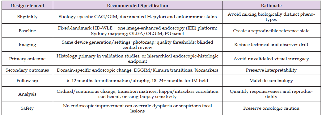
Timing and Analysis: Inflammatory features may change within months, whereas credible assessment of atrophy and especially GIM requires longer follow-up. Six to twelve months may be reasonable for inflammatory and atrophic domains; 18 to 24 months or longer is more defensible for metaplastic field change. Analyses should report continuous or ordinal change, reader agreement, transition matrices and sensitivity analyses for biopsy adequacy, rather than only responder proportions Table 2.
Table 2: Minimum Design Standards for Intervention Trials.
Artificial Intelligence: Useful After Standardization, Not Before It
Artificial intelligence (AI) can support detection, segmentation and automated estimation of EGGIM, and meta-analytic evidence suggests good diagnostic performance for GIM [56,57]. Its most appropriate position in the present framework is within future trial methods and the research agenda. AI may reduce reader variability and quantify field area, but it cannot resolve an undefined endpoint. Algorithms require external validation across devices and populations, locked versions for longitudinal trials, calibration against expert central review and explicit monitoring for temporal or platform drift.
Three validation steps are needed before endoscopic mucosal response can be treated as an independent efficacy endpoint. First, test-retest studies should quantify stability under repeated procedures and readers. Second, longitudinal cohorts should establish responsiveness and the smallest change exceeding measurement error for EGGIM, Kimura-Takemoto categories and mapped field area. Third, trials should determine whether visual change predicts durable histologic or clinical benefit beyond baseline risk stage. The proposed framework also has limitations. Repeated biopsy introduces sampling variability and procedural burden. Platform-specific color signatures may limit generalizability. Regression of precancerous fields is biologically slow and may be confounded by H. pylori eradication or changes in inflammation. Finally, the vitamin B literature is not sufficiently robust to validate the framework by itself. Its value is as a stress test that reveals what future trials must measure more rigorously.
Endoscopic visualization can become a rigorous component of treatment-response assessment in CAG, but only if visual change is defined by domain, measured longitudinally and interpreted with mapped histology. EGGIM and Kimura-Takemoto provide useful starting structures, yet their established role is risk staging rather than validated treatment-response measurement. Pepsinogen and gastrin- 17 add physiologic context but remain supportive. Current vitamin B intervention studies suggest possible benefit in selected populations, particularly for inflammatory or atrophic outcomes, while evidence for reliable reversal of established GIM remains insufficient. Future trials should replace composite effective rates and narrative endoscopy reports with fixed-landmark imaging, blinded central review, lesion-specific follow-up and prespecified concordance rules.
Data sharing not applicable to this article as no datasets were generated or analyzed during the current study.
All authors confirm the absence of any conflict of interest.
This work was supported by the Luzhou Medical Association (Grant No. 2024-YXXM-109).

International Association of Landscape Archaeology, Czech Glass Society, Czech Republic

Department of Chemistry, Semenov Institute of Chemical Physics, USSR Academy of Sciences, Moscow, Russia

Pharmaco-kinetics, dynamics and Drug Metabolism, Touro College of Pharmacy, USA

Professor of Nuclear Medicine, Faculty of Medicine and Surgery, University of Milan, Milan, Italy

Clinical Radiologist (MD) - Department of RADIOLOGY, Cosenza Hospital, Cosenza, Italy





 Abstract
 PDF
PDF