[email protected]
+1 (502) 904-2126
One Westbrook Corporate Center, Suite 300, Westchester, IL 60154, USA
Site Map
 ISSN: 2574 -1241
ISSN: 2574 -1241
ISSN: 2574 -1241
Received: July 30, 2026; Published: August 13, 2026
*Corresponding author: Elnaz Abedini, Faculty of Engineering, Department of Biomedical Engineering, Karabuk University, Karabuk, Turkey
DOI: 10.26717/BJSTR.2026.66.010338
Bone regeneration is an active area that demands the accurate coordination of complicated biological mechanisms. Although classical methods tend to emphasize the osteoinductive or osteoconductive traits of biomaterials, current studies put a highly important emphasis on the interaction between mechanical signals and immune reactions, the so-called immuno-mechanical axis. This review also explores in a detailed manner the recent developments in the field of mechano-immunomodulatory approach to bone regeneration. In particular, the mechanobiological control of the mesenchymal stem cell (MSC) niche, the effect of mechanotransduction pathways, including Piezo1 and YAP/TAZ, and the effect of mechanical substrate stiffness on cell fate are examined in detail. The interactions between the mechanical stimuli on the crosstalk of macrophages (M1/M2) and MSCs, and the immunomodulatory functions of bone healing are described. Moreover, the adoption of artificial intelligence (AI) and machine learning (ML) methods into scaffold design to optimize materials-architecture interactions and predict cell-material interactions is also explained. The latest trends and ideas until 2025, such as digital twins, 4D-bioprinting, and multi-omics AI, are included. Lastly, a solution to translational challenges, especially AI-designed implant regulatory hurdles, and a 2030 roadmap is considered. The review offers an extensive structure on how next-generation biomaterials can be developed to regulate the immuno-mechanical axis of bone regeneration.
Keywords: Bone Regeneration; Mechanobiology; Immunomodulation; Biomaterial Design; Artificial Intelligence; Stem Cell Niche; Macrophage Polarization
Abbreviations: MSC: Mesenchymal Stem Cell; AI: Artificial Intelligence; ML: Machine Learning; BTE: Bone Tissue Engineering; YAP: Yes-Associated Protein; TAZ: Transcriptional Coactivator With Pdz-Binding Motif; ECM: Extracellular Matrix; LPS: Lipopolysaccharide; SVM: Support Vector Machines; ANN: Artificial Neural Networks; GAN: Generative Adversarial Networks; VAE: Variational Autoencoders; CNN: Convolutional Neural Networks; FEA: Finite Element Analysis; RWE: Real-World Evidence; SAMD: Software As A Medical Device; MDR: Medical Device Regulation; TPLC: Total Product Lifecycle; FDA: Food And Drug Administration
Bone tissue is a dynamic system with the ongoing process of remodeling that is governed by a complex of mechanical loading and biochemical signals [1,2]. The topic of bone regeneration is particularly significant in different clinical settings, such as fracture healing, osteoporosis, and repair of defects following tumor resection. Conventional methods in bone tissue engineering (BTE) have been mainly concerned with the development of biomaterials that have osteoconductivity and osteoinductivity features [3]. Nevertheless, their clinical effectiveness has not been very high because of the lack of full knowledge of such a complex biological process as bone healing. Over the last decades, there has been an emergence of the idea of the immuno- mechanical axis, which is the deep and bidirectional connection between mechanical input and immune reactions during bone regeneration [4-6]. This novel paradigm will require that, in addition to biocompatibility of biomaterials, there is a need to focus on the cellular interaction with the mechanical environment and, consequently, the modulation of the cellular response of immune cells.
In bone healing, the immune system is central in all stages. The line of macrophages, in particular, has a wide range of roles, as it triggers the inflammatory reaction and promotes tissue remodeling and repair [7,8]. The phase shift between the pro-inflammatory and the anti-inflammatory/regenerative macrophage M1 and M2 is essential to effective bone regeneration [9,10]. The biochemical factors alone have a significant effect on this phenotypic polarization, and so do the mechanical stimuli to which cells are subjected [11,12]. Indicatively, mechanical signals can have a direct effect on macrophage polarization, and thus, their influence on bone healing, including substrate stiffness, surface topography, and fluid shear forces [13,14]. The other important element in bone regeneration is mesenchymal stem cells (MSCs), which have attracted much attention because of their ability to differentiate into osteogenic and immunomodulatory properties [15,16]. The microenvironment of MSCs is predominantly involved in the tight regulation of their fate and functions [2,17]. The mechanical characteristics of this niche play a ruling role in the proliferation, differentiation, and interaction of MSC with immune cells [18-20]. The main signaling pathways of cellular reactions to mechanical stimuli include mechanotransduction pathways, especially Piezo1 ion channels and YAP/TAZ (Yes-associated protein/ Transcriptional coactivator with PDZ-binding motif) co-activators [21-24]. Not only are these pathways stimulated to promote the osteogenic differentiation of MSCs, but they also influence the immunomodulatory effect of macrophages [25].
The purpose of this review is to integrate the view on the mechano- immunomodulatory strategies in bone regeneration. To start with, the mechanobiological control of the MSC niche and the contribution of mechanotransduction pathways to bone formation will be described. The immunomodulatory processes of the macrophage in response to mechanical stimuli and the crosstalk of the MSCs with the macrophage will be studied further. Third, the deployment of artificial intelligence (AI) and machine learning (ML) algorithms to create streamlined biomaterial scaffolds and forecast cell-material interactions will be mentioned. Lastly, the regulatory issues in the translation of AI-assisted biomaterials to clinical practice and future trends in the topic, specifically in the context of a 2030 roadmap, will be discussed. It is an in-depth analysis that attempts to create new horizons in the creation of improved and focused therapeutic approaches in bone regeneration.
The highly complex bone regeneration process depends on the specific mechanobiological control of the stem cell niche, a dynamic microenvironment determining the growth, differentiation, and motility of the mesenchymal stem cells (MSCs) [2,15]. The niche includes not only physical and chemical properties of the extracellular matrix (ECM), but also the interactions with other cells and biomechanical interactions [17,19]. The mechanical stimuli that the MSCs experience can be of different forms, including substrate stiffness, tension, compression, and fluid shear forces, and these mechanical factors determine the osteogenic differentiation potential of MSCs [1,18,20]. This part will explore the mechanobiological control of the MSC niche, the basic mechanisms behind mechanotransduction, and the importance of substrate stiffness in cell fate determination.
Mechanical Niche and Cell Fate Properties of the MSC
The mechanical properties of the MSC niche form part of the core factors that have a direct effect on the behavior of MSCs in bone-renewal. The stiffness of substrates is a key factor in determining which pathway of osteogenic, chondrogenic, or adipogenic MSCs differentiation takes place [18,21]. As an example, substrates with a stiffness range of values within the physiological range of bone tissue (approximately 25-40 kPa) cause the osteogenic differentiation of MSCs to flourish, whereas softer substrates can stimulate adipogenic or neurogenic differentiation [18,22]. This demonstrates the significance of imitating the mechanical characteristics of the desired tissue in biomaterial development. But, this is not the only thing; how cells detect substrate stiffness and how the cellular responses can be elucidated by a complicated process called mechanotransduction.
It is not just the stiffness of the ECM that affects the mechanical perception of MSCs and, consequently, their further differentiation, but also its topographical properties, porosity, and ligand density [7,19]. Nanostructured surfaces are able to induce osteogenic differentiation faster, improving cell-surface interactions and using certain mechanosensors [7]. These biomimetic strategies hold a great promise in the creation of an optimized biomaterial for bone regeneration. However, contradictory results of various research studies indicate that not all views are completely agreed on the impact of nanostructured cues on M1 polarization [17,22]. This implies that mechanical signal influences on cell behavior are relative to context, and one parameter might not be adequate.
Mechanotransduction Pathways Piezo1 and YAP/TAZ
Mechanotransduction is the way by which mechanical stimuli are translated into biochemical signals in the cells. Although there are many molecular pathways that are implicated, Piezo1 ion channels and YAP/TAZ (Yes-associated proteins/ Transcriptional coactivator with PDZ-binding motifs) co-activators are crucial in the mechanical sensing of MSCs and in bone regeneration [21,23,24]. Piezo1 Ion Channels: Piezo1 is a mechanosensitive cation channel found in the cell membrane. Upon mechanical stress of cells, Piezo1 channels open and cause an influx of calcium ions into the cell [25]. This calcium influx promotes many signaling pathways, which affect cellular processes, especially osteogenic differentiation. Piezo1 has been identified to stimulate osteogenic differentiation of MSCs and accelerate bone formation [13]. Moreover, Piezo1 was reported to be involved in macrophage polarization, which could justify the change to the M2 (regenerative) macrophage phenotype [25]. These results indicate that Piezo1 plays a significant role in the crosstalk between the two MSCs, as well as immune cells, when bone regenerating.
YAP/TAZ Co-activators: YAP and TAZ are important effectors of the Hippo signaling pathway and are important to cellular responses to mechanical stimuli [23,24]. In response to mechanical tension or cells cultivating on solid surfaces, YAP and TAZ relocate to the nucleus and regulate the expression of genes associated with osteogenesis [21,24]. On the other hand, low mechanical tension or soft substrates cause YAP/TAZ to be retained in the cytoplasm and inactivated, thus suppressing osteogenic differentiation [22]. YAP/TAZ activity is tightly connected with cytoskeletal tension and cell shape; increasingly spread and tensed cells tend to have an increased YAP/TAZ activity [21]. This process is the way that the surface topography and stiffness of biomaterials determine the osteogenic fate of MSCs. It is also starting to be evidence-based that YAP/TAZ also contributes to the activities of immune cells and thus augments the molecular underpinning of the immuno-mechanical axis [24].
Effect of Substrate Stiffness on MSC Differentiation
One of the most well-characterized mechanical factors that result in the differentiation pathway of MSCs is substrate stiffness. The in vitro experiments have presented strong evidence to prove that MSCs can be differentiated into different degrees of rigidity [18,21,22]. To illustrate, soft substrates (0.1-1 kPa) facilitate neurogenic differentiation, intermediate stiffness substrates (8-15 kPa) facilitate myogenic differentiation, and stiff substrates (25-40 kPa) facilitate osteogenic differentiation [18]. This effect is used to demonstrate that cells are able to adapt to their surroundings by sensing the mechanical cues and changing their gene expression and cellular behavior in response. Substrate stiffness has been shown to influence MSC differentiation, and these mechanisms happen via cell-ECM interactions and cytoskeletal tension, as well as through mechanotransduction pathways [21]. Cells are able to exert more tension on rigid substrates, which enhances actin-myosin contractility and results in reorganization of the cytoskeleton. This enables the cells that are attached to the ECM through integrins to send mechanical messages to the cell. These cues trigger mechanosensors, such as Piezo1 and YAP/TAZ, which stimulate the transcription of osteogenic transcription factors and, therefore, bone development [23,24].
It is, however, important to note that substrate stiffness does not work by itself in isolation and needs another factor to work; that is, biochemical and biophysical factors. As an example, other factors that cause MSC differentiation include growth factors, cytokines, and cellcell interactions [15]. As such, multi-factorial methods that are based not only on mechanical properties but also biochemical directions need to be embraced when developing biomaterials in bone regeneration. Such a combined method, which is more closely reminiscent of the sophisticated mechanobiological processes of the MSC niche, will allow us to create more powerful methods of bone regeneration. Future studies should focus on the improved comprehension of this sensitive equilibrium amid the mechanical and biochemical cues and their implementation into the biomaterial design.
The complexity of bone regeneration is also contingent upon the fact that it is a complex process that requires the dynamism of the immune system, especially the macrophage [5,6]. The functions of macrophages are wide-ranged since they include the instigation of the inflammatory response, tissue repair, and remodelling after tissue injury [7,8]. In this section, dynamic shifts between the M1 (pro-inflammatory) and M2 (anti-inflammatory/regenerative) macrophage phenotypes, the effect of mechanical stimuli on polarization, and the complex crosstalk between the macrophages and the mesenchymal stem cells (MSCs) are going to be explored.
Macrophage Polarization and its Use in Bone Regeneration
Very flexible immune cells, macrophages may develop many phenotypes upon exposure to microenvironmental cues. The most common phenotypes are the pro-inflammatory M1 macrophages and the anti- inflammatory/regenerative M2 macrophages [4,26]. M1 Macrophages are normally triggered by pro-inflammatory cytokines, including lipopolysaccharide (LPS) and interferon-gamma (IFN-γ). Producing significant levels of pro-inflammatory cytokines (such as TNF-α, IL-1β, IL-6) and reactive oxygen species (ROS), M1 macrophages are engaged in host defense against pathogens as well as in the removal of damaged tissues [27]. The arrival of M1 macrophages is one of the steps that must be taken during the first bone healing process to induce the inflammatory response and to clear out the damaged tissue. Still, an extended or excessive M1 reaction might lead to persistent inflammation and bone resorption, therefore impairing regeneration [28]. Surprisingly, other research shows that M1 macrophages might induce osteogenic differentiation of MSCs indirectly, even if this effect is typically transient and circumstances dependent [27].
Usually stimulated by anti-inflammatory cytokines, including IL-4 and IL-13, M2 macrophages, by creating anti-inflammatory cytokines (e.g., IL-10, TGF-β) and growth factors (e.g., VEGF, PDGF), M2 macrophages aid tissue repair, angiogenesis, and immunosuppression [29]. Solving inflammation and starting tissue repair, the M1-M2 transition is critical in the last stages of bone healing. Directly promoting osteoblast differentiation and bone formation are M2 macrophages [9,29]. Therefore, a compelling method to maximize bone healing [4,6] is the development of biomaterials that drive macrophage polarity towards M2 phenotype.
Mechanical Stimuli Effect on Macrophage Polarization
Biochemical signals are not the only factors that control macrophage polarization, and mechanical stimuli to which the cells are exposed are heavily regulated as well. The phenotyping and functions of macrophages can be affected by certain factors, including substrate rigidity, surface topography, shear forces in fluid, and cyclic mechanical forces [11-14]. Substrate Stiffness: Macrophage response to substrate stiffness has been found to be comparable to the MSCs’ response, with a few differences. In general, the softer substrates are more prone to M2 polarization, and harder substrates could stimulate M1 polarization [12]. Nevertheless, the complication of such an association is obvious in contrasting findings provided by various bodies of knowledge. In the case of a few studies, it is proposed that the stiffness of nanostructured surfaces causes M1 polarization [17], whereas it is also proposed that certain stiffness ranges favor M2 polarization [12]. These inconsistencies demonstrate the necessity of a better insight into the effect of particular geometries, chemical functionalizations, and experimental environments of nanostructured cues on macrophage responses.
Mechanotransduction Pathways: The Piezo1 ion channels have a major role in macrophage mechanosensing and polarization. Mechanical stress on Piezo1-activated macrophages stimulates calcium influx, promoting M2 polarization and TGF-β1 release, which subsequently promotes bone formation [25]. Equally, the alternating mechanical loading has the capability of activating the other (M2-like) macrophages, thus facilitating osteogenesis [9,14]. The results imply that the mechanical characteristics of biomaterials may directly mediate the immunomodulatory functions of macrophages and direct bone regeneration.
Macrophage-MSC Crosstalk and Mechanical Control
The interaction between macrophages and MSCs crosstalk is important in bone regeneration, and this is heavily controlled by mechanical signals [10,16]. MSCs may secrete several factors which determine the polarization of macrophages (e.g., PGE2), and macrophages may secrete cytokines and growth factors which determine osteogenic differentiation of MSC [30,31]. Effects of MSCs on the polarization of the Macrophage: MSCs can induce an M2 macrophage polarization in macrophages through their immunomodulatory activity. This action may take place by secretion of soluble factors like prostaglandin E2 (PGE2) [30]. PGE2 facilitates M2 polarization through changes in the metabolic condition of the macrophage. Thus, the development of biomass to increase the immunomodulatory properties of MSCs is one of the possible solutions that can maximize bone regeneration. Macrophage effects on MSC Differentiation. Macrophages and especially M2 macrophages can directly promote osteogenic differentiation and bone formation in MSCs. As an example, TGF-β1 released by M2 macrophages is a significant growth factor, which facilitates osteogenic differentiation of MSCs [29]. Moreover, the conditional media of mechanically stimulated macrophages have been indicated to stimulate osteogenic genes in human mesenchymal stromal cells [31]. Such results have shown that the impact of the mechanical stimulus on macrophages, in turn, can indirectly affect the osteogenic fate of MSCs.
Mecano-Immunomodulatory Biomaterials. To achieve success in bone regeneration, the mechanical sensing of MSCs and the immunomodulatory action of the macrophages should work together [3,4,6]. These biomaterials have the opportunity to maximize their physical characteristics, including surface topography, stiffness, and porosity, to induce macrophage polarization to M2 phenotype and also induce MSC osteogenic differentiation [7,32]. As an illustration, BCP ceramics stimulated in certain micro-vibration fields have proven to be effective with cascade amplification on immune activation up to bone regeneration [32]. Such a combined strategy has potential in the formulation of more potent therapeutic programs through manipulation of the immuno-mechanical axis in bone regeneration.
Conventional trial-and-error methods of designing biomaterials to regenerate bones are lengthy and expensive processes that are, in most cases, unable to sufficiently consider the multifactoriality of the complex biological systems. The recent progress in artificial intelligence (AI) and machine learning (ML) provides the opportunity to redefine the design of biomaterials, such that the optimization of the scaffold architecture and enhanced predictability of the cell-material interactions can be achieved [33-35]. In this section, the contribution of ML and generative AI to the optimization of scaffold architecture, predicting cell-material interactions, and the future of next-generation biomaterials to regenerate bone will be discussed.
Scaffold Architecture Optimization Using Machine Learning
The architecture of scaffolds plays a significant role in the bone regeneration of biomaterials. The pore size, pore interconnectivity, surface area, and mechanical properties are the parameters that have a direct effect on cell adhesion, proliferation, differentiation, and nutrient/ waste transport [36]. ML algorithms have the potential to train on large datasets and learn these complex interactions to generate an optimal scaffold architecture to obtain desired biological behavior. Supervised Learning Approaches: Supervised learning models are used to discover the relationships between known scaffold designs and their experimentally measured biological responses (e.g., osteogenic differentiation, immunomodulation). As an example, artificial neural networks (ANNs) or support vector machines (SVMs) can be trained to estimate the behavior of various scaffold geometries and different material mixes on cell behaviors [37]. These models can be used to determine the best scaffold parameters, which can be used in a particular application by utilizing the information obtained through high-throughput screening data. To illustrate this, a single study came up with an explainable ML-based probabilistic framework to design scaffolds in bone tissue engineering [33].
Generative Design and Generative AI: Generative AI provides the capability to design new and optimized scaffolds using existing data. Such tricks as generative adversarial networks (GANs) and variational autoencoders (VAEs) are able to produce novel scaffold geometries that fulfill given design constraints and goals (e.g., a specific mechanical stiffness and porosity) [38]. These techniques are very useful in coverage of design space and the identification of new scaffold designs that would not be feasible using conventional techniques. The example is that AI-optimized lattice structures can be employed in biomechanical scaffold design [36]. It has also been shown that AI-assisted designing of tissue engineering scaffolds with the help of virtual tomography and 3D convolutional neural networks (CNNs) [37].
Multi-omics AI and Cell-Material Interactions Prediction
Successful bone regeneration is dependent on the interaction of biomaterials with the cells. Such interactions depend on a variety of parameters, such as the surface chemistry of the material, its topography, mechanical characteristics, as well as the genetic and epigenetic conditions of the cells. These complicated interactions can be predicted using ML models, and the biological responses of the biomaterials can be predicted.
1. Cell-Material Interaction Prediction: ML algorithms can be trained on materials datasets (e.g., stiffness, roughness, chemical composition) and cellular outputs (e.g., gene expression, protein production, differentiation status) datasets. These models have the ability to forecast the possible biological performance of innovative biomaterials without any significant in vitro or in vivo tests [39]. This hastens research and development activities and allows more specific biomaterial designs. Indicatively, biomimetic nanofiber scaffolds can reduce the inflammatory response, which can be predicted using machine learning and graph theory to regenerate tissues [39].
2. Multi-omics AI: It is one of the recent trends as of 2025 to combine multi-omics data (genomics, transcriptomics, proteomics, metabolomics) with AI. It is an effective method to study the molecular complexity of cell-material interactions. Multi-omics artificial intelligence can be used to investigate the impact of biomaterials on cells in a comprehensive manner, to identify new biomarkers, and to develop individualized bone regeneration plans. Indicatively, AI has been applied to optimize PCL/PEG electrospun scaffolds to promote in vivo wound healing [40].
4D Bioprinting and Digital Twins
The future of AI-based biomaterial design is in the fact that it will be integrated with new technologies, including digital twins and 4D bioprinting.
1. Digital twins: Digital twins are virtual versions of a physical system or process—biomaterial design. In biomaterial design, a digital twin of a scaffold can be designed to simulate the material properties, scaffold architecture, and predicted biological responses. These computer models are constantly revised and optimized by real-life data, and it is possible to optimally tune the behavior of scaffolds in real-time and change it. By shortening the time and cost of the development process, digital twins can minimize the in vitro and in vivo experiments.
2. 4D Bioprinting: Although 3D bioprinting uses fixed structures, 4D bioprinting can be used to print smart biomaterials capable of deforming or altering their functionality with time. These biomaterials have the capability of changing in a pre-programmed way in reaction to external factors like temperature, pH, light, or mechanical stress. The design of 4D bioprinted scaffolds can be optimized using AI, and structures that are capable of responding dynamically to physiological loads and better supporting bone regeneration can be produced. This prepares the customized and responsive therapeutic modalities in bone regeneration.
Overall, AI and ML are solving the shortcomings of existing methods in the design of biomaterials, thus allowing the creation of optimized scaffold designs, predicting cell-scaffold interactions, and combining them with new technology, like digital twins or 4D bioprinting. These developments present an optimistic avenue for creating more efficient and specific therapy techniques in bone regeneration.
Although artificial intelligence (AI) and machine learning (ML)- based biomaterials have transformative potential in the area of bone regeneration, there are serious problems with the implementation of these new technologies in the clinic. In particular, the regulatory approval, the safety, the efficacy, and the ethical issues related to AI-designed implants are the main obstacles that may stall the development of this sphere [41,42]. In this section, the regulatory environment of AI-enabled medical equipment, the difficulties it faces, and a roadmap for hastening the rate of translational advancements in this area by 2030 shall be outlined.
Regulatory Environment of AI-Medical Devices
The use of AI and ML algorithms is becoming part of the design, production, and utilization of medical equipment. But as opposed to the traditional medical devices, AI devices can be adaptive and continuously learning systems, which challenge the established regulatory frameworks in a unique way [43,44]. Regulatory agencies are actively attempting to keep up with these new technologies based in various territories of the globe (e.g., US FDA, EU MDR, China NMPA, etc.) [45,46]. USUS Food and Drug Administration (FDA): The FDA has been at the forefront of regulating medical devices based on AI/ML. These devices are usually regarded by the FDA as “Software as a Medical Device” (SaMD), and the FDA has embraced a Total Product Lifecycle (TPLC) model [47]. The purpose of this approach is to make the device safe and effective during its lifecycle and take into account the fact that it is able to learn and change even after it is released into the market. Nevertheless, there are still ambiguities concerning the way to deal with the constant change of algorithms and performance [48]. In high-risk fields like cardiovascular devices, AI/ML-enabled device usage must undergo a stringent assessment procedure [48].
European Union Medical Device Regulation (MDR): EU MDR has put tighter controls on medical devices, although it is relevant to AI-enabled devices [42]. The MDR requires conformity tests depending on the risk of the device. It may also be difficult to collect clinical evidence and conduct conformity assessment using AI algorithms due to their complexities and lack of transparency, particularly those that are highly risky [42]. Analysis of the regulatory environment within particular regions, including the role of AI in imaging, identifies gaps in the regulatory environment and the requirements for the future [49]. China National Medical Products Administration (NMPA): China is also actively engaged in the regulation of AI medical devices and is building up its own model. In comparison with the USUS and the EU, comparative research points to the various regulatory strategies and their effects on the world market [45,50].
Regulatory Challenges Experienced
The regulatory approval of AI-powered biomaterials and implants has a number of distinct challenges:
• Transparency and Explainability: AI algorithms have black-box decision-making processes, which are not explained. Regulatory organizations need to be informed as to the reason some device decisions are made or why performance is attained in a given manner. This is highly essential to patient safety [44]. Although explainable AI (XAI) algorithms are being devised to resolve this problem, they are not yet at a stage of full development to use in clinical settings [33].
• Data Quality and Bias: AI is as good as the data that it is trained on. The lack, absence, or incompleteness of data may create mistakes in the functioning of the devices with AI or inequality in some groups of patients [51]. The regulatory authorities are delegating tougher standards so as to ensure the representativeness, quality, and diversity of the datasets being utilized.
• Continuous Learning and Adaptation: AI/ML can learn indefinitely by using real-world information and enhance its performance. Nevertheless, a major challenge of this adaptation is control under the regulation to provide safety and efficiency of the device. Approval of each update to the algorithm would be a drag on innovation. As a consequence, terms like change management and the application of the Real-World Evidence (RWE) are getting more and more significant [47].
• Cybersecurity and Privacy: Doctors can extract a lot of sensitive information related to patients using AI-enabled medical equipment. This data is highly sensitive, and the security and privacy of this data against cyberattacks and data breaches are major concerns of regulatory bodies. Moreover, poor use or control of devices is also dangerous.
• Ethical Concerns: AI-enabled medical devices also present ethical issues about responsibility, accountability, and patient autonomy [52]. The problem of accountability in the event of an error (manufacturer, physician, AI algorithm) is even more complicated with the active usage of such technologies.
2030 Roadmap: Translational Progress: Accelerating Regulatory entities need to be resolved to achieve their potential in bone regeneration involving AI-enabled biomaterials, and translational gains need to be fast-tracked. The actions that can be undertaken in this sector by 2030 are as follows:
• International Harmonization and Collaboration: The gaps between the regulatory structures in various states make it difficult to enter the international market. The increased cooperation and integration of regulatory authorities can help to approve AI-enabled medical devices faster and more efficiently [46,53].
• Creation of Transparency and Explainability Standards: Industry/Academic Standards, common standards on improving the transparency and explainability of AI algorithms should be created. This will enable regulatory bodies to be in a position to evaluate the safety and effectiveness of devices better. Elucidation ML-based frameworks are going in this direction strongly [33].
• Real-World Evidence (RWE) and Continuous Performance Monitoring: In order to deal with the constant learning capability of AI-enabled gadgets, it is important to consider the incorporation of real-world evidence (RWE) into regulatory procedures. Monitoring of the performance of the devices has to be done continuously after the release into the market, and the updates should be recorded [47].
• Education and Capacity Building: Education and capacity building are required on AI and ML technologies among regulatory authorities, healthcare providers, and biomaterial developers. This will see the establishment of knowledge and the safe execution of new technologies.
• Enhancement of Ethical Frameworks: Broad ethical frameworks are to be established and put in place to provide the ethical utilization of AI-enabled medical devices. Such frameworks are expected to deal with the problem of responsibility, fairness, privacy, and patient autonomy [52].
• In Silico Clinical Trials: Computational modeling and simulations (in silico clinical trials) can be an integral part of regulatory procedures, since they can speed the process of developing and approving AI-enabled biomaterials [54]. Such methods can minimize the quantity of experimental tests so as to minimize the cost and speed up the innovation [55-60].
In conclusion, in order to achieve clinical success of AI-enabled bio-material in the sphere of bone regeneration, scientific and new technology advancement must be supported by a powerful and active control system, transparency criteria, and ethics. The 2030 roadmap will eliminate these challenges through a multidisciplinary approach to AI in bone regeneration (Figures 1-3, Tables 1 & 2).
Figure 1

Table 1: Correlation of Biomechanical Parameters with Immune and Osteogenic Outcomes.
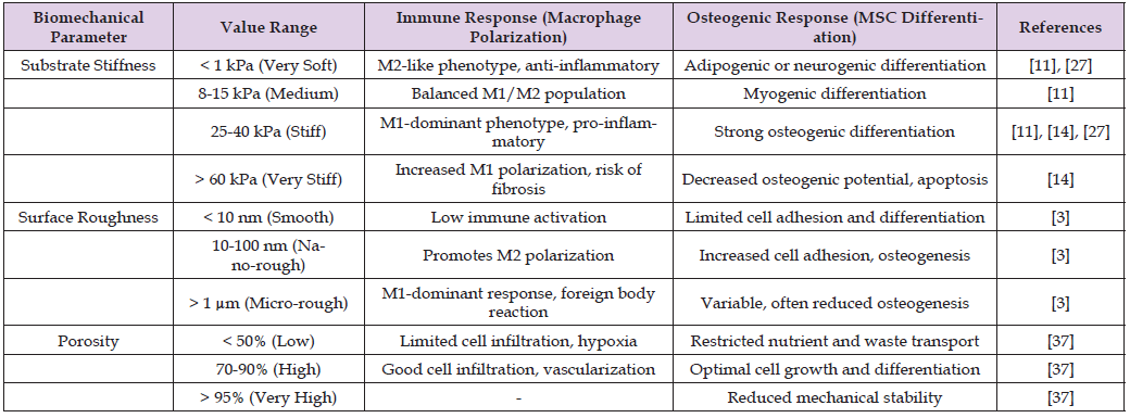
Figure2
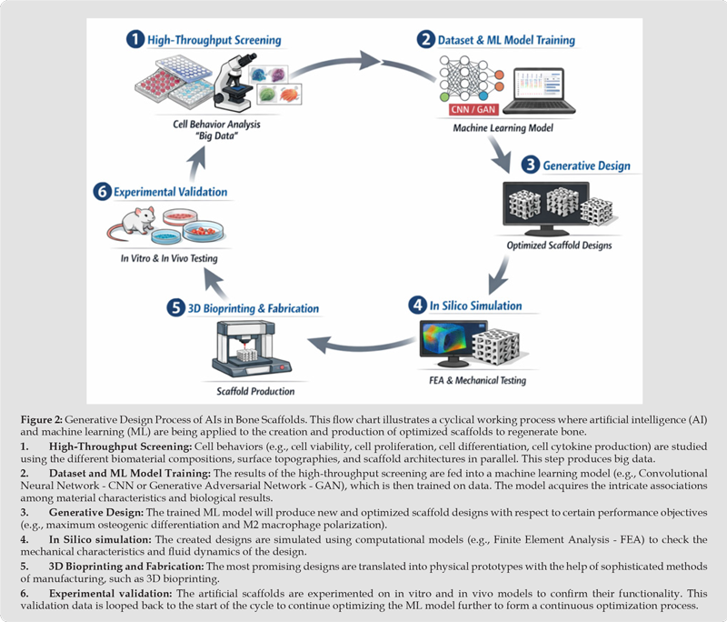
Table 2: Comparison of AI Models for Scaffold Design Optimization.

Figure 3


International Association of Landscape Archaeology, Czech Glass Society, Czech Republic

Department of Chemistry, Semenov Institute of Chemical Physics, USSR Academy of Sciences, Moscow, Russia
Associate Professor at Department of Breast and Thyorid Surgey, Chongqing General Hospital, China

Professor of Nuclear Medicine, Faculty of Medicine and Surgery, University of Milan, Milan, Italy

Department of Pharmaceutical Sciences, South University School of Pharmacy, USA





 Abstract
 PDF
PDF