[email protected]
+1 (502) 904-2126
One Westbrook Corporate Center, Suite 300, Westchester, IL 60154, USA
Site Map
 ISSN: 2574 -1241
ISSN: 2574 -1241
ISSN: 2574 -1241
Received: March 31, 2026; Published: April 14, 2026
*Corresponding author: Anna Piro, Consiglio Nazionale delle Ricerche, Istituto di Bioimmagini e Sistemi Biologici Complessi, Sede Secondaria, Via Tommaso Campanella 115 88100 Catanzaro, Italy
DOI: 10.26717/BJSTR.2026.65.010173
In a selected group of male patients affect by cardiologic defects and undergoing to digoxin therapy, color vision resulted to be a useful biologic marker to identify this drug intoxication influencing retina and the brain color vision pathways
Keywords: Color Vision; Drug Intoxication; Retina Structure; Retina Cells Function; Digoxin
Na, K-ATPase are energy consuming ion pumps that are required for maintaining ion homeostasis in most cells. In the retina, Na, K-ATPase are especially important to sustain the dark current in photoreceptor cells needed for rapid hyperpolarization of rods and cones in light. Cardiac glycosides like digoxin inhibit the activity of Na, K-ATPase by targeting their catalytic alpha subunits. This leads to a disturbed ion balance, which can affect cellular function and survival. Digoxin induced cell death specifically in photoreceptor cells with no or only minor effects in other retinal cell types. Degeneration was restricted to photoreceptors of the central retina, depended not by the presence of rhodopsin, activated stress signaling, and induced genes involved in degeneration, inflammation and oxidative stress.
Synaptic interactions to extract information about wavelenght, and thus color, begin in the retina with three classes of light-sensitive cells: rod photoreceptors at low light levels, multiple types of cone photoreceptors that vary in spectral sensitivity, and intrinsically photosensitive ganglion cells that contain the photopigment melanopsin. The mechanisms that underlie the perception of color have interested scientists since the 17th century. Sir Isaac Newton recognized that “The rays, to speak properly, are ot colored. In them there is nothing else than a certain power and disposition to stir up a sensation or that color”. We now recognize that “stirring up a sensation” for the perception of color arises from complex neural computations implemented in the multistage process that begins with the distinct spectral tuning properties of cone photoreceptors and then proceeds through the retinal circuitry on to the lateral geniculate nucleus, the primary visual cortex. So, an intrinsic process which drives the eyes to the human brain. Color can be defined as occupying a three-dimensional color space with three principal maxes: red versus green, blue versus yellow and white versus black. The red/green and blue/yellow axes define the hue or chromaticity, whereas the white/black axis defines luminace in humans [1].
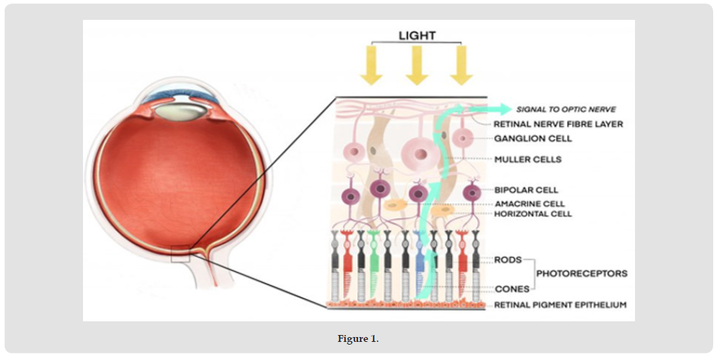
Altough the transmission channels along which nervous impulses are conducted are well established anatomically as separate units, knowledge of the way in which each of the retinal structures contributes to the totals and the mechanisms whereby signals are transferred from cell to cell, are less clear at present. Two main features of the organisation of the retina are central to color processing, the functional linkage between cells (coupling or convergence) laterally through the receptive field, laterally through the receptive field, and the feedback mechanism which horizontal cells mediate either electronically or chemically to influence the final output of the cone receptors. The former is the anatomical basis for spatial summation and the latter for lateral inhibition, both of which are found frequently in sensory pathways. These features have a far greater consequence than mere economic channeling of responses, resulting in the cells losing their independence as single units; they greatly influence the sensitivity of the eye and the response to edges or demarcations of stimuli. On the deficit side the coupling means the spatial resolution is reduced. The cell dendrites of horizontal cells make contact with cone receptors at their bases or pedicles.
Two types of horizontal cells have been identified [2] and these were originally thought to control the rod and cone signals respectively, but both types only make dendritic contact with cones. The difference between horizontal cell types is indicated by the numbers of cones they serve, one type typically contacting seven cones and others twelve cones. Near the fovea each cone is probably in contact with at least is probably in contact with at least four horizontal cells and one foveal horizontal cell connects between seven and twelve cones, depending on its type. Towards the periphery the number of cones to horizontal cells increases to between thirty and forty; in this way they extend lateral coverage, summating or gathering information over a wide area. Horizontal cells also link up with other horizontal cells, and they can also “drive” or stimulate into action, bipolar cells and through them ganglion cells. The lateral interactions ensure that receptors outside the dendritic spread of a bipolar cell can communicate easily. All neurones transfer their signals to adjacent neurones through pharmacological agents, the neurotransmitters contained within synaptic vesicles
There exist a wide group of color vision disturbance which are acquired during life, predominantly the result of ocular or general disease, the consequence of exposure to a chemical, toxin or medication, or resulting from physical injury to the head. The incidence of such disturbances is at least 5% [3]. It is realised that colour vision changes provide a valuable means of monitoring the progress of a disease, or the toxic effects of a chemical substance, whether exposure is deliberate for therapeutic purposes or unintentional in the case of an industrial hazard. The effectiveness of treatment can occasionally be assessed by the continued monitoring of recovery of an acquired colour vision disturbance. A wide variety of chemicals and drugs affect colour vision indirectly, usually as a consequence of damage to the retina and/or optic nerve inflammation and atrophy of the optic nerve is a frequent consequence of long-term medication of a great many drugs, notably the anti-inflammatory agents used to treat cardiac agents.
Cardiac glycosides, principally digitalis and the associated drugs digitoxin or digoxin which are widely used to increase cardiac output, or to correct arrhythmias, have been known for some time to affect colour vision at therapeutic levels. The complaint of chromatopsias, frequently xanthopsia is common, objects appearing tinged with yellow, blue, green or red; blue halos can appear around lights [4] found a colour vision disturbance in 80% of toxified patients. The prolonged action of digoxin and digitoxin gives rise to the most pronounced colour perception disturbances, altough even these are generally reversible. A close correlation between the extent of colour vision disturbance and serum digoxin concentration is reported [4]. Optic or retrobulbar neuritis is the underlying cause for colour vision disturbance, with prominent macular cone damage; central scotoma are thus typical. Zanen [5] proposed that the site of disturbance is retinal. Cozijnsen and Pinckers [6], Verriest [7] noted a red-green disturbances in cases of severe intoxication; Grutzner [8,9], Babel and Stangos [10] report a blue-like defect. Towbin [11] found a significantly sensitivity to green. A specific disturbances of the Stiles red mechanism was observed by Gibson [12]. Their patient showed an inherited deutereranomalous defect, with a superimposed protan and tritan involvement as a consequence of the drug toxicity. Alken [13] confirmed protan-like defects in patients taking single 1 mg doses of digoxin, which had earlier been reported by Cozijnsen and Pinckers [6] in digoxin intoxicated patients. Repeated daily doses of 0.375 to 0.75 mg for two to three revealed green disturbances in his sample confirming the general findings of Towbin [11].
We investigated 16 cardiac male patients hospitalized in Istituto Nazionale per il Ricovero e Cura degli Anziani (INRCA) from Cosenza province (Calabria, Southern Italy). Obviously, this choosing allowed us to avoid Lyon genetic phenomenon showed in the females where the compensation X chromosome phenomenon is present. In our work, 8 out 16 patients showed normal color vision; 7 out 16 patients showed color vision deficiency; 1 out 16 patients showed red-green inherited colorblindness. The age onset of the cardiac disease is 30 months (range 0,2-72 months); New York Association Class mean was II (range I-III); digoxin treatment mean time was 53 months (range 2-144 months). Color vision deficiency in 7 out 16 patients were subdivided as follows: left eye, 2 patients showed light protan/deutan color vision deficiency on red/green axis; 1 patient showed great protan/deutan color vision deficiency on red/green axis; 3 patients showed light tritan color vision deficiency on blue/yellow axis. Regarding the right eye, we showed 4 patients with normal color vision; 2 patients showed light tritan color vision deficiency on blue/yellow axis; 1 patient showed great protan/deutan color vision deficiency on red/green axis. We excluded inherited colorblind patient from the analysis of the results. 8 patients showing normal color vision were treated with digoxin with a mean time of 4 years than the group of patients showing color vision deficiency (Figure 1).
Figure 1
We confirm the important and precise color vision role as a useful biological and physiological marker to identify the digoxin drug toxicity from the retina to the color vision brain pathways.
Authors thank Fondazione Cassa di Risparmio di Calabria e Lucania for its contribution.

International Association of Landscape Archaeology, Czech Glass Society, Czech Republic

Department of Chemistry, Semenov Institute of Chemical Physics, USSR Academy of Sciences, Moscow, Russia

Pharmaco-kinetics, dynamics and Drug Metabolism, Touro College of Pharmacy, USA

Professor of Nuclear Medicine, Faculty of Medicine and Surgery, University of Milan, Milan, Italy

Clinical Radiologist (MD) - Department of RADIOLOGY, Cosenza Hospital, Cosenza, Italy





 Abstract
 PDF
PDF